| # | key | img1.png | img2.png | txt |
|---|
1
|
iu_xray__00000000
|
|
|
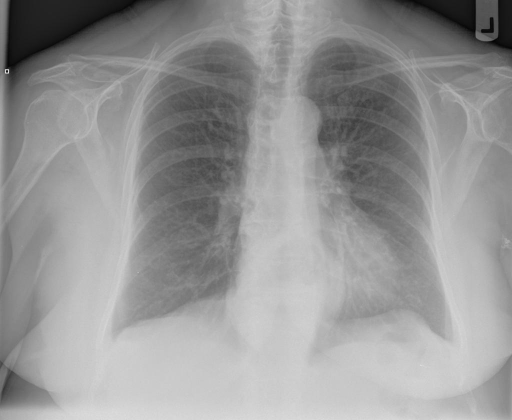
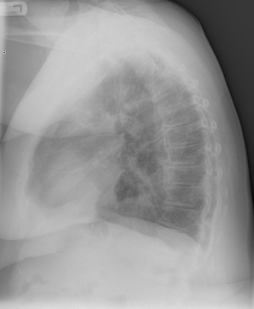
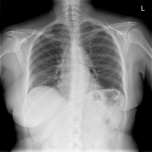
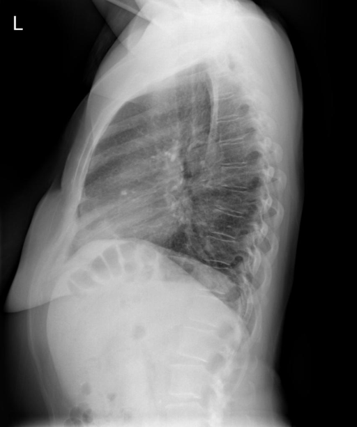
user: Take a look at these two images: <|img1|> and <|img2|>. Describe them and provide a diagnosis.
assistant: The chest X-ray images are of good quality with appropriate patient positioning, showing both posteroanterior (PA) and lateral views. The heart size and pulmonary vascularity are within normal limits. A large hiatal hernia is evident, seen as a soft tissue density mass behind the heart. The lung fields are clear with no signs of focal airspace disease, pneumothorax, or pleural effusion. Degenerative changes are noted in the thoracic spine, likely indicative of osteoarthritic changes. The diaphragms are in normal position without any subdiaphragmatic free air, and the costophrenic angles are sharp. The airways appear unremarkable with no signs of hilar enlargement or adenopathy. Bony structures, including the ribs and clavicles, are intact with no fractures or lytic lesions visible. The cardiomediastinal silhouette is normal, and both lung hila are in typical position without vascular congestion.
|
2
|
iu_xray__00000001
|
|
|
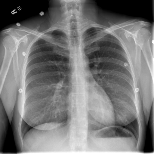
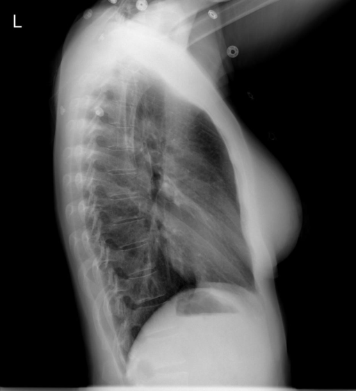
user: Here are two images: <|img1|> and <|img2|>. Explain their contents and share your diagnosis.
assistant: The provided chest X-rays include both a posteroanterior (PA) and a lateral view, with well-defined image quality and proper patient positioning. The airway is central with no signs of displacement. The bony structures, including ribs and clavicles, appear intact without evidence of fractures. The cardiac and mediastinal contours are normal, showing no enlargement or abnormal shadowing. The diaphragm is well-positioned, with clear costophrenic and cardiophrenic angles, indicating no pleural effusions. Lung parenchyma is clear in all visualized zones, without evidence of focal consolidations, vascular prominence, or alveolar air space disease. Hilar structures appear normal with no signs of adenopathy. The patient's age and sex are not specified, but these findings are consistent with a healthy chest X-ray.
|
3
|
iu_xray__00000002
|
|
|
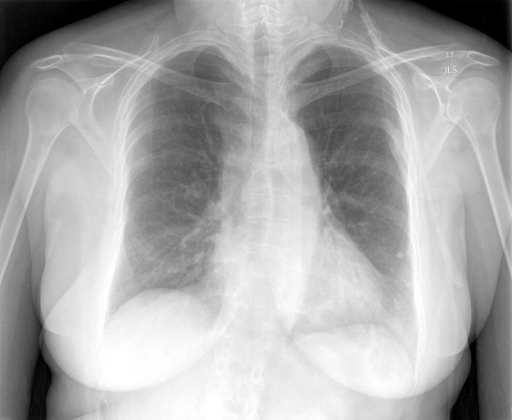
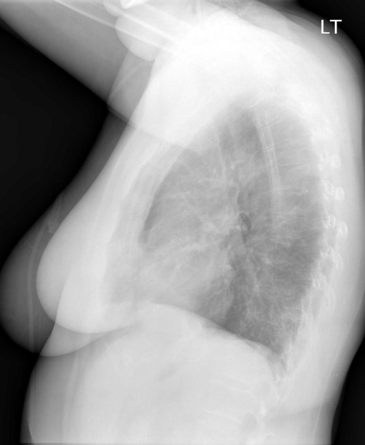
user: Here are two images: <|img1|> and <|img2|>. Explain their contents and share your diagnosis.
assistant: The chest X-rays provided are of acceptable quality with standard positioning, one being an anteroposterior (AP) view and the other a lateral view. The image demonstrates a left lower lobe calcified granuloma, suggesting a prior granulomatous infection. The heart size appears within normal limits with no evidence of cardiomegaly. There is no visible pleural effusion or pneumothorax. The right lung displays mild medial atelectasis. There are signs of mild emphysema, indicated by the increased radiolucency and possibly flat diaphragms. The bones and breast shadows are unremarkable, with no fractures or lytic lesions observed. The costophrenic angles are clear, and there is no free air under the diaphragm. The hilar region is not enlarged, and vascular markings appear within normal limits. These findings are documented without reference to patient demographics as they were not provided.
|
4
|
iu_xray__00000003
|
|
|
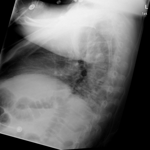
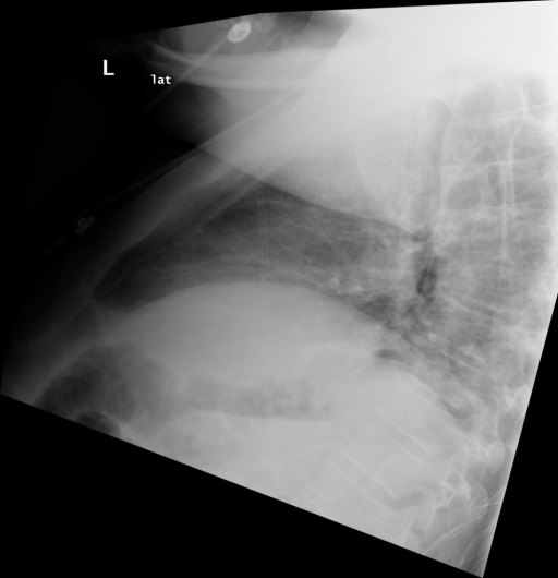
user: The following images are given: <|img1|> and <|img2|>. Describe them and provide a diagnosis.
assistant: The images provided are lateral chest X-rays. The quality and patient positioning appear appropriate for evaluation. The airways are clear without evidence of hilar adenopathy or enlargement. Examination of bones reveals a displaced fracture of the mid third of the right clavicle. The cardiomediastinal silhouette is normal in size and contour. Diaphragmatic contours are intact with no signs of free air beneath. The pleural edges show no signs of pneumothorax or significant pleural thickening. Lung fields exhibit bilateral opacities noted at the lung bases. No definitive pleural effusion observed. Hilar structures on the visible side are within normal limits. Patient demographics such as age and sex are not provided.
|
5
|
iu_xray__00000004
|
|
|
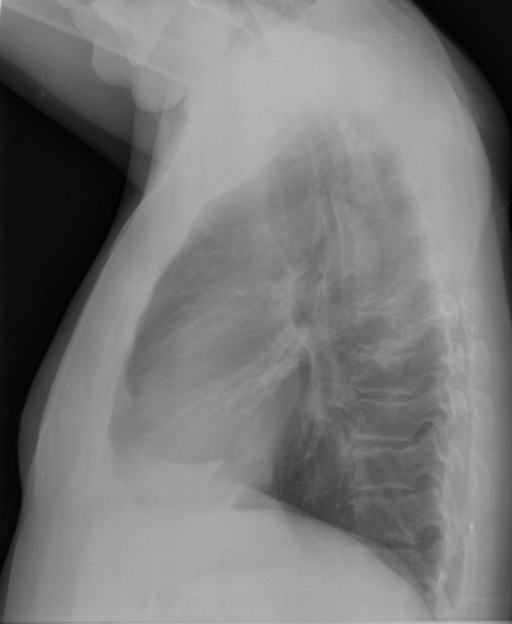
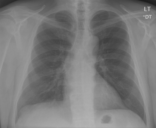
user: Here are two images: <|img1|> and <|img2|>. Explain their contents and share your diagnosis.
assistant: The chest X-ray images provided include both lateral and posteroanterior (PA) views. The images are of good quality with appropriate contrast and positioning, allowing for clear assessment of the thoracic structures.

Airways: The trachea is midline with no signs of hilar enlargement or any abnormalities.

Breast shadows and Bones: The osseous structures, including ribs and spine, appear intact with no evidence of fractures, lytic lesions, or crowding.

Cardiomediastinal contour: The cardiac silhouette is normal in size and shape, with no apparent abnormalities in the mediastinal region. Costophrenic angles are sharp, indicating no pleural effusions.

Diaphragm: Both hemidiaphragms are well-positioned with no sign of free air beneath them, suggesting the absence of abdominal perforation.

Edges and Extrathoracic tissues: There is no evidence of pneumothorax, pleural thickening, or apical fibrosis. The soft tissues are unremarkable.

Fields: Lung parenchyma is clear bilaterally across the upper, middle, and lower zones. There is no evidence of alveolar consolidation, vascular prominence, or plural effusions.

Hilum: The hila appear normal with standard anatomical positioning. No abnormal enlargement of vessels or lymph nodes is evident.

The images show no acute pathology and present as unremarkable.
|
6
|
iu_xray__00000005
|
|
|
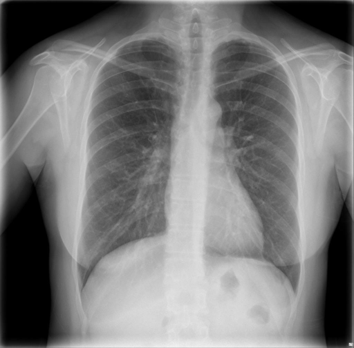
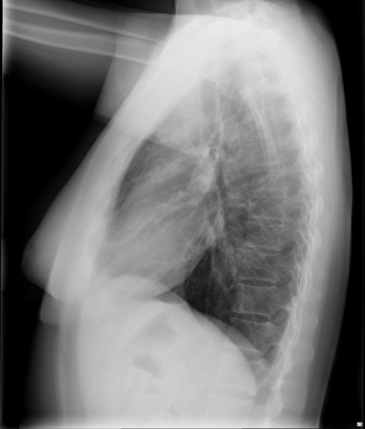
user: Examine the following images: <|img1|> and <|img2|>. Describe what you observe and offer your diagnosis.
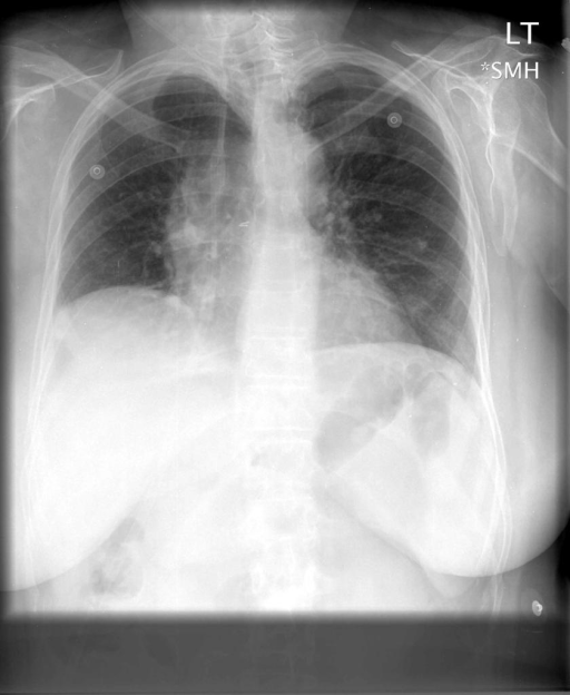
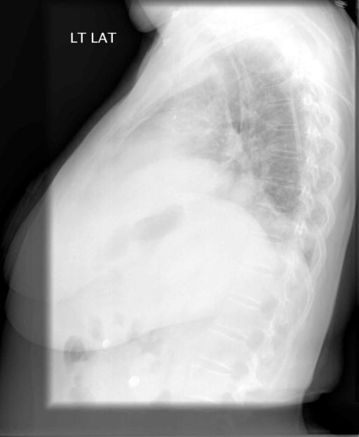
assistant: The chest X-ray images provided include both posteroanterior (PA) and lateral views. The image quality is adequate with correct patient positioning and appropriate contrast.

Airways are clear with no signs of hilar adenopathy or enlargement. The bones, including ribs and clavicles, show no fractures or lytic lesions. The cardiac silhouette appears normal in size, and the mediastinum is unremarkable without noticeable shifts. The hemidiaphragms are properly positioned without free air present underneath.

There is evidence of biapical scarring, though the rest of the lung fields are clear, with no signs of alveolar air space disease or pleural effusions. The pleural edges show no pneumothorax or significant pleural thickening. The hila are in a normal position with no abnormal vessel prominence.

Patient demographic details such as age and sex are not provided. The findings suggest stability with noted biapical scarring as the sole abnormality.
|
7
|
iu_xray__00000006
|
|
|
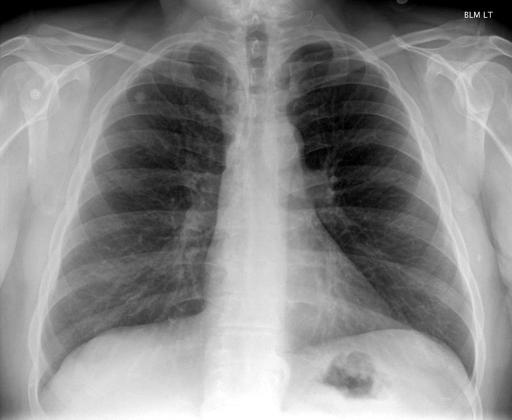
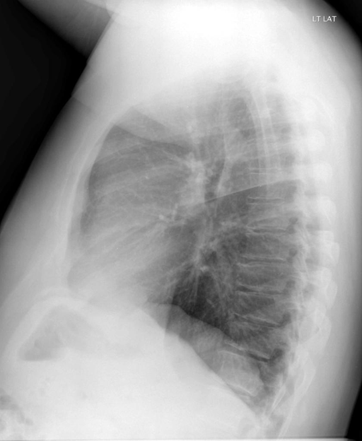
user: Examine the following images: <|img1|> and <|img2|>. Describe what you observe and offer your diagnosis.
assistant: The two chest X-rays include a posteroanterior (PA) and lateral view, both of high quality with appropriate patient positioning and adequate image contrast. Clinical evaluation of the airways reveals a midline trachea with no signs of hilar adenopathy or enlargement. Breast shadows and bones are normal, with a noted syndesmophyte suggesting possible spinal ankylosis or an inflammatory process. There are no rib fractures or lytic lesions evident. The cardiomediastinal contour presents a normal heart size, and no abnormalities are noted in the mediastinum or costophrenic angles, confirming absence of pleural effusion. Diaphragms are well-positioned with no free air underneath, ruling out pneumoperitoneum. Edges of the lungs demonstrate no evidence of pneumothorax or pleural thickening. Examination of the lung fields shows clear parenchyma with bilateral symmetry and no signs of air space disease. Both hilum appear to be in normal position with no vascular abnormalities. Notably, an 11 mm well-circumscribed nodule in the right upper lobe remains unchanged from a previous evaluation 7 years ago, indicating stability. No demographic details are provided for age or sex.
|
8
|
iu_xray__00000007
|
|
|
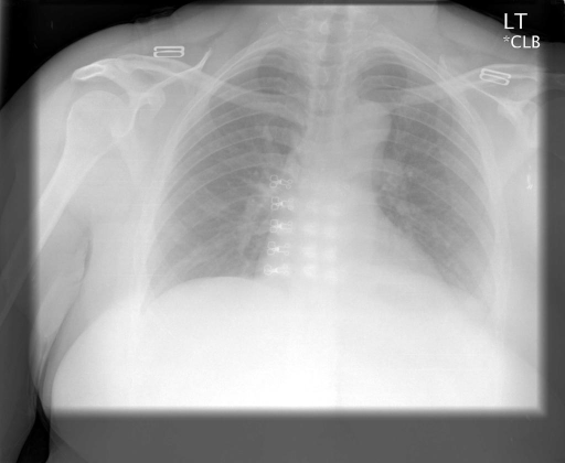
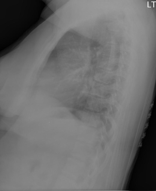
user: Review these images: <|img1|> and <|img2|>. Describe what you see and provide your diagnosis.
assistant: The chest X-ray images are presented with acceptable quality, showing a frontal and lateral view. The patient is positioned correctly, most likely upright, allowing for comprehensive examination of thoracic structures.

Airways: The trachea is midline, with no evidence of hilar adenopathy or enlargement.

Breast shadows and Bones: No rib fractures or lytic bone lesions are observed. There is slight rib crowding consistent with reduced lung volumes.

Cardiomediastinal Contour: The cardiac silhouette and mediastinal contours appear within normal limits.

Diaphragm: The hemidiaphragms are appropriately positioned with no free air detected under the diaphragm, ruling out perforation of abdominal organs.

Edges: The lateral lung margins show no signs of pneumothorax or pleural thickening. Extrathoracic tissues appear unremarkable.

Fields: The lung parenchyma is clear, with no alveolar airspace disease. There is vascular crowding noted due to low lung volumes, but no pleural effusions are detected.

Hilum: The hila are in a normal position, and vascular structures appear intact without distortion.

The patient is of unknown age and sex based on the available images and description.
|
9
|
iu_xray__00000008
|
|
|
user: Examine the following images: <|img1|> and <|img2|>. Describe what you observe and offer your diagnosis.
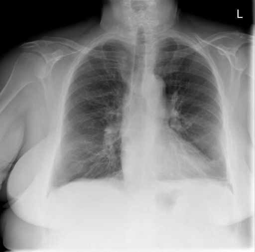
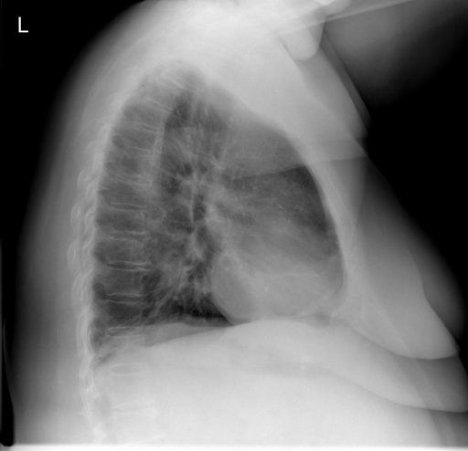
assistant: The provided chest X-rays include both a posteroanterior (PA) and lateral view. The images are of good quality, with appropriate patient positioning and contrast.

Airways appear normal without evidence of hilar adenopathy or enlargement. Assessment of the bones shows no rib fractures, lytic lesions, or crowding. The cardiac silhouette is normal, and the pulmonary vascularity is within normal limits. There are no pleural effusions, as the costophrenic angles are clear. The diaphragms are in normal position without free air underneath. The lung edges reveal no pneumothorax, pleural thickening, or fibrosis. Lung fields are well-aerated with mild bibasilar atelectasis. Minimal atherosclerotic changes are noted in the thoracic aorta. The hilum shows normal positioning and vascular structures.

All observations should be correlated with clinical findings for completeness.
|
10
|
iu_xray__00000009
|
|
|
user: Review these images: <|img1|> and <|img2|>. Describe what you see and provide your diagnosis.
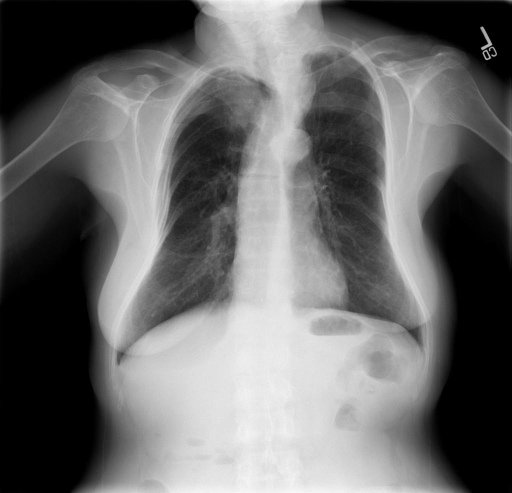
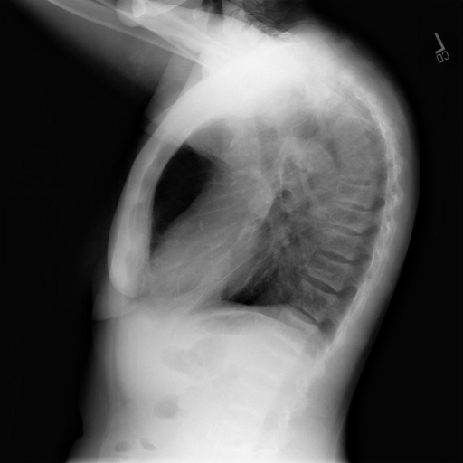
assistant: The X-ray images are of standard quality and positioning, featuring both posteroanterior (PA) and lateral views of the chest. The trachea is midline, indicating no apparent shift. Examination of the breast shadows and bony structures reveals no rib fractures or lytic bone lesions, while the superior thoracic spine remains unchanged from prior evaluations.

The cardiomediastinal silhouette appears normal with no evidence of cardiac enlargement or deviation. The diaphragms are properly positioned, with no free air detected beneath them. The lung fields display lucency consistent with emphysema, showing no significant progression or change from previous assessments. No pneumothorax is evident, and there are no signs of focal airspace disease or pleural effusion.

The hilum appears unremarkable with no abnormalities in vessel positioning or structure. A vague density in the medial right lung apex is stable, likely due to overlapping bony structures rather than a pathologic finding.

This description applies to an adult patient, though specific demographic details such as age and sex are not provided.
|
11
|
iu_xray__00000010
|
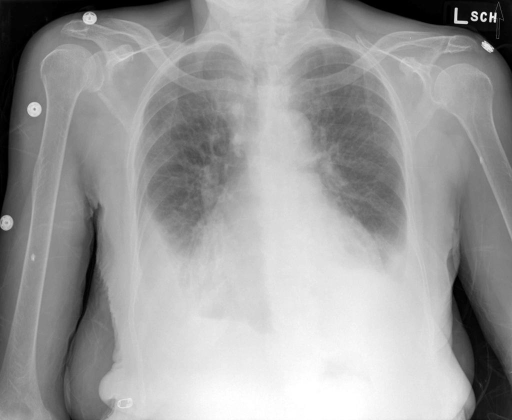
|
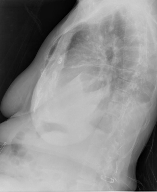
|
user: Examine the following images: <|img1|> and <|img2|>. Describe what you observe and offer your diagnosis.
assistant: The series consists of a frontal and lateral chest X-ray. The images are of good quality with appropriate contrast and show the patient in the upright position.

Airways: There are no abnormalities in the trachea or bronchi, and no evidence of hilar adenopathy is observed.

Breast shadows and Bones: No rib fractures or bony lesions are present. The skeletal structures appear normal, with no rib crowding observed.

Cardiomediastinal Contour: The cardiac silhouette is enlarged, suggesting possible cardiomegaly, but the mediastinal contour remains within normal limits. No abnormalities are seen within the central structures, and the costophrenic angles show blunting, more pronounced on the left, which might suggest an underlying pleural effusion.

Diaphragm: The hemidiaphragms are in a normal position without evidence of free intraperitoneal air.

Edges: No signs of pneumothorax or pleural thickening are visible. The apices and extra-thoracic tissues are unremarkable.

Fields: The lung parenchyma is clear of nodules or masses. Bilateral interstitial opacities are noted, which may indicate interstitial lung disease. Stable-appearing right perihilar calcified granulomas are present, suggesting a history of granulomatous disease.

Hilum: The hila appear in a normal position and are not enlarged.

Patient demographics such as age and sex are not provided.

In summary, the X-ray reveals cardiomegaly, bilateral costophrenic angle blunting, and interstitial opacities with stable calcified granulomas. No acute pathologies such as pneumothorax or free air under the diaphragm are observed.
|
12
|
iu_xray__00000011
|
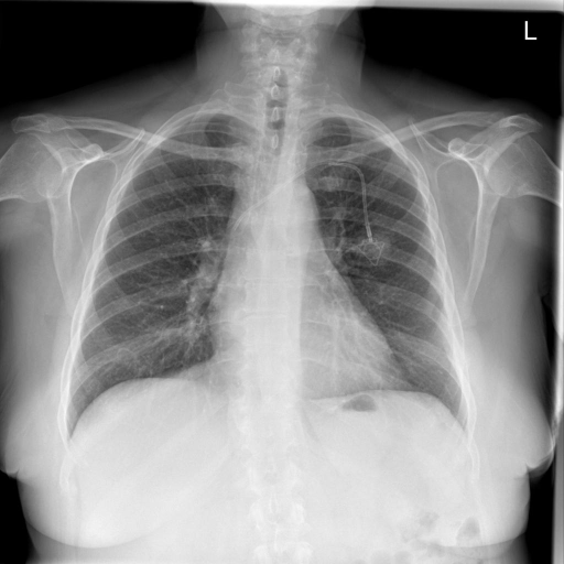
|
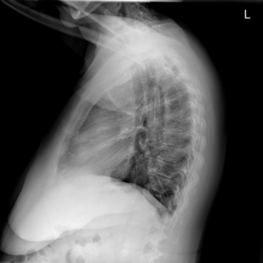
|
user: Take a look at these two images: <|img1|> and <|img2|>. Describe them and provide a diagnosis.
assistant: The chest X-ray images display a posteroanterior (PA) and lateral view of the chest. The image quality is adequate with proper patient positioning.

The airways are clear without signs of hilar adenopathy or enlargement. Examination of the breast shadows and bones reveals no rib fractures or lytic bone lesions. Degenerative changes are noted in the thoracic spine, but no rib crowding is present.

The cardiomediastinal contour is normal; the heart size is within normal limits. There are no pleural effusions, and the costophrenic angles are clear.

The diaphragm is positioned appropriately with no free air observed beneath it.

No pneumothorax or pleural thickening is visible in the peripheral edges. The lung fields appear clear in the upper, mid, and lower zones bilaterally, with no alveolar air space disease or vascular prominence.

The left XXXX appears stable with the tip approximating the high superior vena cava. The hilum shows no abnormalities in the position or vessels.

Patient details such as age and sex are unspecified.
|
13
|
iu_xray__00000012
|
|
|
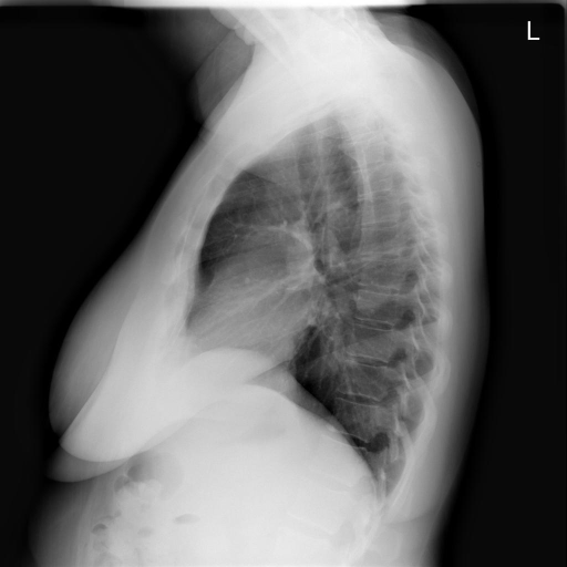
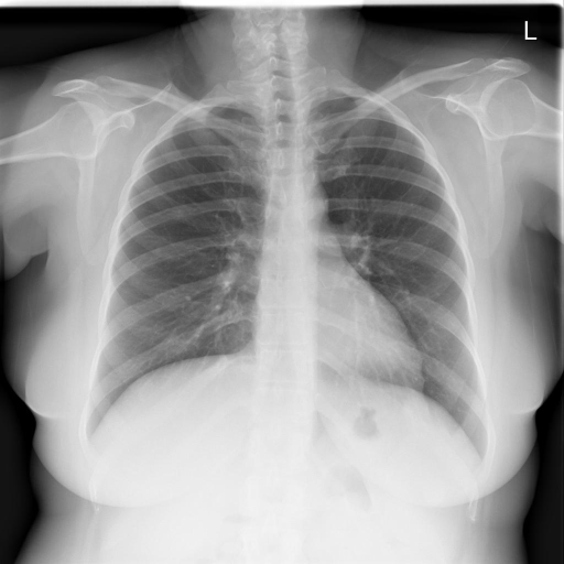
user: Consider the images <|img1|> and <|img2|>. Describe your observations and state your diagnosis.
assistant: The chest X-ray images include a posteroanterior (PA) and lateral view, showing no positioning errors and good image quality.

Airways: The trachea is midline with no signs of shift or hilar enlargement.

Breast shadows and Bones: The osseous structures, including ribs and clavicles, show no fractures or lytic lesions. No rib crowding indicating volume loss is observed.

Cardiomediastinal contour: The cardiac silhouette is normal in size, and the mediastinal contours are clear. There's no widening or abnormal contour. Costophrenic angles are sharp, with no evidence of pleural effusion.

Diaphragm: Both hemidiaphragms are in a normal position without elevated or flattened areas. No free subdiaphragmatic air is present.

Edges: There is no evidence of pneumothorax or pleural thickening. The apices and lateral edges show no abnormalities.

Fields: The lung parenchyma appears clear in the upper, mid, and lower zones bilaterally, without signs of airspace disease or vascular prominence.

Hilum: The hila are in a normal position with no abnormal vascular markings or expansion seen.

These observations indicate a normal chest X-ray for the specific demographic presented, showing no pathological findings such as focal airspace disease, pleural effusion, or pneumothorax.
|
14
|
iu_xray__00000013
|
|
|
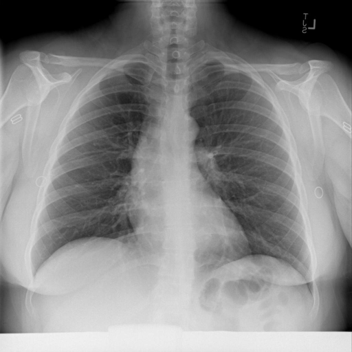
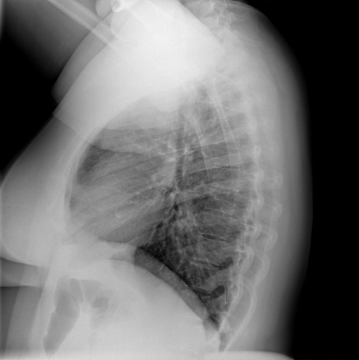
user: The following images are given: <|img1|> and <|img2|>. Describe them and provide a diagnosis.
assistant: The chest X-ray images provided include both an anteroposterior (AP) and a lateral view. The image quality is sufficient for evaluation, with correct patient positioning and appropriate contrast.

Airways: The trachea appears central without signs of deviation or obstruction. No hilar enlargement is noted.

Breast shadows and Bones: The bone structures, including the ribs, scapulae, and clavicles, appear intact without evidence of fractures or lytic lesions. Mild scoliosis is observed in the spine.

Cardiomediastinal Contour: The cardiac silhouette is normal in size and shape, and the mediastinum is unremarkable. There are no signs of cardiomegaly. The costophrenic angles are sharp without evidence of pleural effusion.

Diaphragm: Both hemidiaphragms are in a normal position with no free air indicating viscus perforation.

Edges: The pleural spaces show no signs of pneumothorax or significant thickening. The apices and lateral edges are clear of any pathological findings.

Fields: The lung parenchyma is clear with no evidence of alveolar infiltrates, consolidations, or vascular prominence. The lung fields show good inflation and symmetry across both sides.

Hilum: The hilar regions are normal without evidence of lymphadenopathy or vascular abnormalities.

In summary, the X-ray images reveal clear lungs with no indications of pleural effusion, pneumothorax, or significant cardiac or mediastinal abnormalities. Mild scoliosis of the spine is noted, but no acute bony or parenchymal pathology is present.
|
15
|
iu_xray__00000014
|
|
|
user: Two images are provided: <|img1|> and <|img2|>. Analyze them and provide your diagnosis.
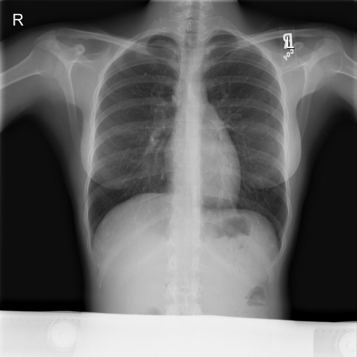
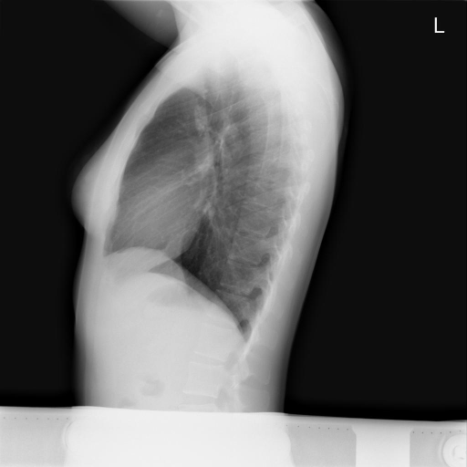
assistant: The chest X-rays show good image quality with appropriate positioning in both posteroanterior (PA) and lateral views, allowing for clear evaluation of thoracic structures.

Airways appear normal without evidence of hilar adenopathy or enlargement. The bones, including ribs and clavicles, are intact with no visible fractures or lesions. The cardiomediastinal contour shows the heart size within normal limits and no abnormalities in the silhouette or chamber size. The costophrenic angles are clear without pleural effusion.

The diaphragms are well-positioned with no signs of free air beneath. Lung edges reveal no pneumothorax or pleural thickening. The lung fields are clear with no focal alveolar consolidation, signs of pulmonary edema, or vascular prominence, confirming symmetric air distribution across lobes.

The right upper lung features a dense nodule, potentially indicating a previous granulomatous process, along with observed mediastinal calcification. The hilum presents a normal position without vessel enlargement or abnormal junctions.

Patient demographic information, age and sex, was not provided. The findings support a stable chest condition without acute pathologies.
|
16
|
iu_xray__00000015
|
|
|
user: The following images are given: <|img1|> and <|img2|>. Describe them and provide a diagnosis.
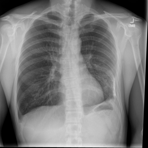
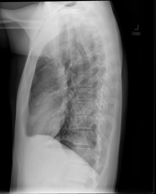
assistant: The chest X-rays consist of a posteroanterior (PA) and lateral view. The images demonstrate good quality with proper patient positioning, and the contrast is appropriate for evaluation.

Airways: The trachea is midline with no evidence of hilar adenopathy or enlargement.

Bones and Breast Shadows: Bones appear intact with no rib fractures or lytic lesions observed. Breast shadows are symmetrical.

Cardiomediastinal Contour: The cardiac silhouette is within normal limits, and the mediastinal contours appear unremarkable. The costophrenic angles are sharp without evidence of pleural effusion.

Diaphragm: Hemidiaphragms are in their appropriate positions, with no free air observed beneath them.

Edges: No evidence of pneumothorax, pleural thickening, or fibrosis. The thoracic soft tissues are unremarkable.

Fields: The lung parenchyma is clear with no focal air space opacities. Pulmonary vascularity is normal. A calcified opacity is noted at the left lung base, suggestive of an old calcified granuloma possibly due to previous infection like tuberculosis or resolved empyema.

Hilum: Normal position and appearance of the hilar structures with appropriately sized pulmonary vessels.

Patient demographic details such as age and sex are not provided.
|
17
|
iu_xray__00000016
|
|
|
user: Review these images: <|img1|> and <|img2|>. Describe what you see and provide your diagnosis.
assistant: The chest X-ray consists of both frontal and lateral views, displaying adequate quality and appropriate positioning. The cardiomediastinal silhouette appears unchanged with an unfolded aorta. There is a streaky opacity at the left base likely indicating scarring or discoid atelectasis. A small calcified granuloma is present in the mid-right lung. Additionally, small nodular opacities are observed, projecting over the right base in the right costophrenic sulcus, near the posterior right ninth rib, and over the anterior T10 vertebral body. No focal airspace consolidation or pleural effusion is evident. The diaphragmatic and hilar regions appear unremarkable in these images. The patient's age and sex are not specified in the description.
|
18
|
iu_xray__00000017
|
|
|
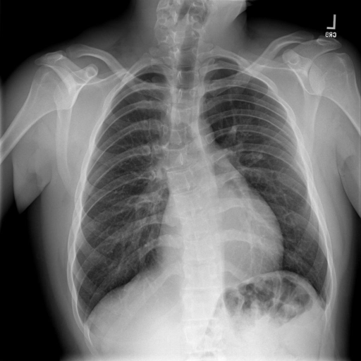
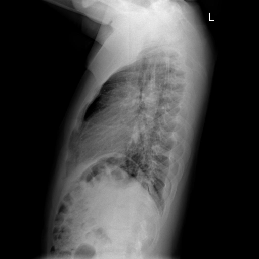
user: Given the two images <|img1|> and <|img2|>, describe their contents and give your diagnosis.
assistant: The chest X-ray images include an anteroposterior (AP) and lateral view. The positioning appears adequate, with appropriate contrast and detail for clinical assessment.

In the AP view, there is evident dextroscoliosis of the thoracic spine. The airways show no abnormalities, with clear hilar regions. The bony structures, including the ribs, do not exhibit fractures or lytic lesions. The cardiomediastinal contour is normal, with no cardiomegaly, and the cardiac silhouette is well-defined. The costophrenic angles appear sharp without signs of pleural effusion.

Both hemidiaphragms are appropriately positioned, and there is no subdiaphragmatic free air, suggesting no diaphragmatic or abdominal issues. There is no indication of pneumothorax or pleural thickening along the lung edges. The lung fields are symmetrical, with no signs of consolidation, vascular prominence, or alveolar infiltrates observed.

The hila are unremarkable, with visible pulmonary vessels in a normal arrangement. The images do not disclose the patient's age or sex, which would aid in further context, though these findings suggest normal pulmonary and cardiac features aside from the noted spinal curvature.
|
19
|
iu_xray__00000018
|
|
|
user: Take a look at these two images: <|img1|> and <|img2|>. Describe them and provide a diagnosis.
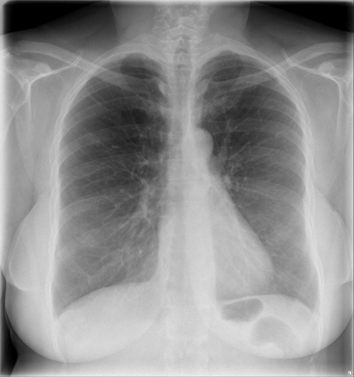
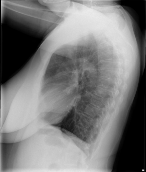
assistant: The chest X-rays display both posteroanterior (PA) and lateral views of high image quality, with appropriate patient positioning and contrast.

Airways appear normal, with no evidence of hilar adenopathy or enlargement. The bone structures, including ribs and spine, show no fractures or lytic lesions. Cardiomediastinal contours are well-defined, with a normal heart size. The mediastinum is appropriately positioned, and the costophrenic angles appear minimally blunted, which might suggest a small pleural reaction without overt effusion.

The diaphragm is intact, with no free air visible under either hemidiaphragm. Lung fields, divided into upper, mid, and lower zones, show clear parenchyma without focal airspace consolidation, pleural effusion, or pneumothorax. Apices appear normal with no signs of pleural thickening or fibrosis.

The hilum maintains a normal appearance with appropriately positioned vasculature and no evidence of vascular congestion or abnormal enlargement of the pulmonary vessels.

Overall, the findings indicate normal heart and mediastinal structures with no significant acute pathology visible in the lungs or pleural spaces.
|
20
|
iu_xray__00000019
|
|
|
user: Review these images: <|img1|> and <|img2|>. Describe what you see and provide your diagnosis.
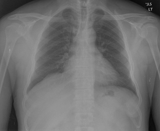
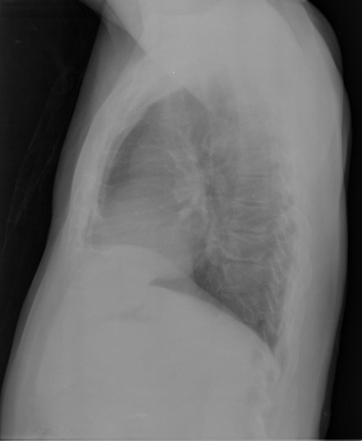
assistant: The chest X-ray images include both anteroposterior (AP) and lateral views, showing adequate contrast and proper patient positioning. The airways appear clear with no signs of hilar adenopathy. Breast shadows are not prominent, and the osseous structures, including the ribs and thoracic vertebrae, show no fractures or lytic lesions indicative of acute abnormalities. The cardiomediastinal silhouette is normal, with no enlargement of the heart or mediastinal structures, and the costophrenic angles are sharp, indicating no pleural effusion. The diaphragms are well-positioned, with no evidence of free air beneath them. The lung fields are clear, showing no signs of focal consolidation, pneumothorax, or fibrosis. The hila are in their normal positions without any vascular enlargement. Patient demographic information is not provided.
|
21
|
iu_xray__00000020
|
|
|
user: Two images are provided: <|img1|> and <|img2|>. Analyze them and provide your diagnosis.
assistant: The provided chest X-ray images include a posteroanterior (PA) and a lateral view, exhibiting appropriate patient positioning and satisfactory image quality with adequate contrast.

Airways are clear with no evidence of hilar adenopathy or enlargement. There are no visible fractures or lesions in the ribs or chest bones, and no sign of rib crowding, indicating no notable volume loss. The cardiomediastinal contour is normal, with a cardiac silhouette of appropriate size and no abnormalities in the central structures. The costophrenic angles are sharp, with no pleural effusions noted.

The diaphragms are well-positioned, with no free air detected under the diaphragm. The lung edges show no signs of pneumothorax, pleural thickening, or fibrosis. Examination of the lung fields reveals no focal consolidation, suspicious pulmonary opacities, or vascular prominence, and both lungs appear clear and symmetrical across upper, mid, and lower zones.

The hila position appears normal, and vessels are unremarkable with no anomalies identified. Based on these observations, the chest X-ray findings are within normal limits.
|
22
|
iu_xray__00000021
|
|
|
user: Consider the images <|img1|> and <|img2|>. Describe your observations and state your diagnosis.
assistant: The chest X-ray images provided are in both posteroanterior (PA) and lateral views. The image quality is adequate with appropriate contrast and patient positioning.

Airways: The trachea and main bronchi appear normal without evidence of hilar adenopathy or airway enlargement.

Breast shadows and Bones: The ribs and clavicles display normal alignment with no visible fractures. There are severe arthritic changes noted at the acromioclavicular joints, with mild arthritic changes observed in the thoracic spine.

Cardiomediastinal contour: The heart is not significantly enlarged, and the cardiac silhouette is within normal limits. The aorta shows atherosclerotic changes. Costophrenic angles are clear indicating no pleural effusions.

Diaphragm: The diaphragms are well-positioned with no evidence of free air underneath them.

Edges: There are no signs of pneumothorax, and the pleural edges are smooth without thickening or plaques.

Fields: The lung parenchyma is clear in all zones—upper, mid, and lower—bilaterally, with no signs of alveolar air space disease or vascular prominence.

Hilum: The pulmonary vessels appear normal, and the positional relationships between the superior pulmonary vein and inferior pulmonary artery are preserved.

The patient appears to be an older adult, but age and sex specifics are not provided in the initial details.
|
23
|
iu_xray__00000022
|
|
|
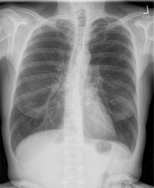
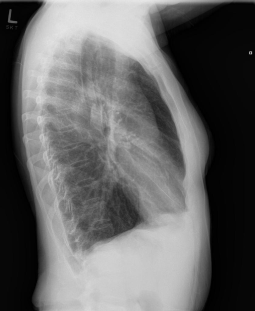
user: Examine the following images: <|img1|> and <|img2|>. Describe what you observe and offer your diagnosis.
assistant: The chest X-ray images provided show a PA and lateral view. The image quality is adequate with correct patient positioning and appropriate contrast.

Airways are patent without evidence of hilar adenopathy or enlargement. The breast shadows are unremarkable, and bony structures show no fractures or notable anomalies. The ribs are intact without crowding. The cardiomediastinal contour is normal with an appropriately sized cardiac silhouette, and mediastinal structures appear unremarkable. No abnormalities are noted in the costophrenic angles.

The diaphragm is well positioned with clear definition of both hemidiaphragms. There is no free air under the diaphragm.

Edge findings reveal no pneumothorax or pleural thickening. The pleura and apices are clear, with no plaques or fibrosis noted. Extrathoracic tissues are unremarkable.

Lung fields are hyperexpanded, indicating possible obstructive lung disease; however, they show no airspace consolidation, masses, or nodules. There is no evidence of pleural effusion. The pulmonary vessels and hilum are within normal limits, with no displacement or abnormal junctions noted.

Overall, the patient appears to have hyperexpanded lungs without focal consolidation, pleural effusion, pneumothorax, or cardiomediastinal abnormalities. The visualized bones and soft tissues are normal.
|
24
|
iu_xray__00000023
|
|
|
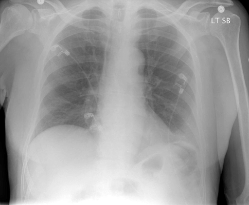
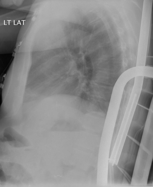
user: You’re given two images: <|img1|> and <|img2|>. Analyze them and provide a diagnosis.
assistant: These chest X-rays include an anteroposterior (AP) view and a lateral view. The images are of good quality with proper contrast, allowing for a clear assessment of anatomical structures.

Airways: The trachea appears midline with no evidence of hilar adenopathy or enlargement.

Breast shadows and Bones: The visualized ribs and clavicles show no fractures or lytic lesions. The bony structures are intact with no rib crowding, indicating no evidence of volume loss.

Cardiomediastinal contour: The cardiac silhouette is normal in size and shape. The mediastinum is appropriately positioned with defined central structures. The costophrenic angles are sharp without pleural effusions noted.

Diaphragm: The diaphragms are well-positioned with no free air under the diaphragm, suggesting no perforation of abdominal viscera.

Edges: No pneumothorax is detected. The pleura appear normal with no thickening, plaques, or apical abnormalities. Extrathoracic soft tissues show no anomalies.

Fields: Lung fields are clear bilaterally across upper, mid, and lower zones with no evidence of alveolar air space disease, vascular prominence, pleural effusion, or consolidation.

Hilum: The hila are in normal position with standard anatomical characteristics. There is no abnormality in the vessels or structures at the hilum.

Patient demographic information, such as age and sex, is not provided and thus not included in this description.

In conclusion, the chest X-rays are unremarkable with normal findings across all examined areas.
|
25
|
iu_xray__00000024
|
|
|
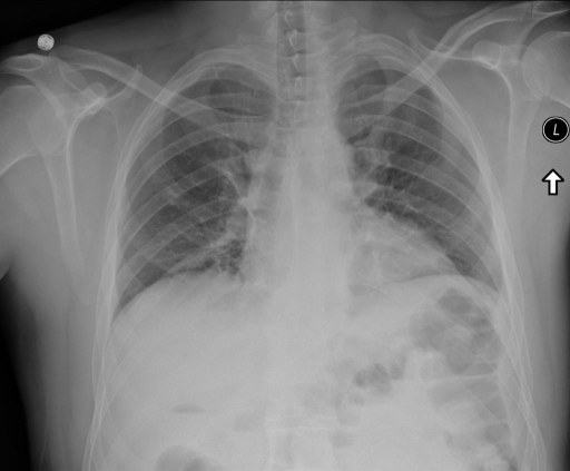
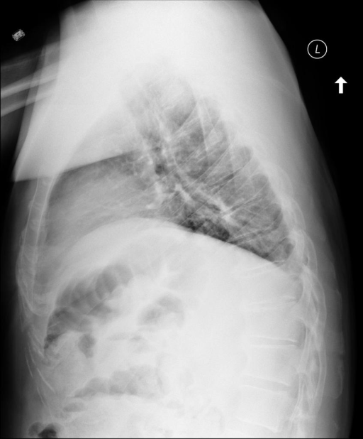
user: Given the two images <|img1|> and <|img2|>, describe their contents and give your diagnosis.
assistant: The chest X-rays provided include both anteroposterior (AP) and lateral views. Image quality appears adequate, with proper contrast and positioning.

Airways: The trachea is midline with no evident deviation or enlargement of the hilar structures.

Breast shadows and Bones: No fractures or lytic bone lesions are present. The ribs appear intact without evidence of crowding.

Cardiomediastinal contour: The cardiac silhouette is normal in size. Pulmonary vascularity appears within normal limits. The costophrenic angles are sharp and clear, indicating no pleural effusion.

Diaphragm: The hemidiaphragms appear at normal heights with no evidence of free air suggestive of perforation.

Edges: No pneumothorax or pleural thickening is detected.

Fields: There is bilateral lung volume loss. Bandlike opacities are observed in the right lung, consistent with atelectasis. No alveolar air space disease or pleural effusions are present.

Hilum: The hila are positioned normally with no significant vascular enlargement.

The patient information (age and sex) was not available. This description provides a thorough assessment of the X-ray findings for use in pre-training a multimodal language model.
|
26
|
iu_xray__00000025
|
|
|
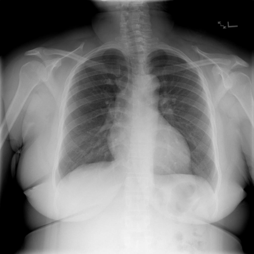
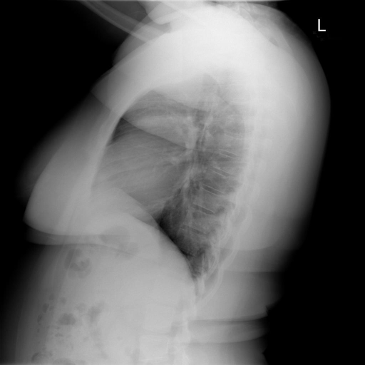
user: Given the two images <|img1|> and <|img2|>, describe their contents and give your diagnosis.
assistant: The images include a PA and lateral view chest X-ray of satisfactory quality, indicating proper patient positioning and adequate contrast for assessment. The clinical observations are as follows:

Airways are patent with no visible hilar enlargement or adenopathy. Breast shadows and bones appear normal, with no evidence of rib fractures or bone lesions. Cardiomedial contour is normal, the heart is of appropriate size and shape, and there are no abnormalities noted in the central structures or costophrenic angles. The diaphragms are properly positioned with no free air observed beneath them.

Edges are clear with no signs of pneumothorax, pleural thickening, or pleural plaques. Lung fields across all zones are clear and symmetrical without any alveolar airspace disease, infiltrates, or vascular congestion. The hilar region shows typical anatomy with normal positioning of the superior pulmonary vein and inferior pulmonary artery.

The images correspond to an adult patient with no specific sex or age provided.
|
27
|
iu_xray__00000026
|
|
|
user: Consider the images <|img1|> and <|img2|>. Describe your observations and state your diagnosis.
assistant: The chest X-rays presented are of adequate quality with proper patient positioning, including anterior-posterior and lateral views.

Airways: The trachea is midline with no apparent hilar enlargement.

Breast shadows and Bones: No rib fractures or lytic bone lesions are observed. Bone structures appear intact without evidence of abnormalities.

Cardiomediastinal contour: The cardiac silhouette is within normal limits, showing stability compared to prior imaging. The mediastinal area is clear, and surgical clips are present, indicating previous procedures. No abnormalities in the costophrenic angles suggest an absence of pleural effusion.

Diaphragm: The hemidiaphragms appear at normal levels with no evidence of free air indicating perforation.

Edges: No evidence of pneumothorax, apical fibrosis, or pleural thickening is noted.

Fields: The lung fields are clear in all zones with no signs of focal airspace disease or vascular anomalies.

Hilum: The hilar regions are unremarkable, with normal positioning and no signs of vascular congestion.

The patient's age and sex are not provided. Overall, the X-ray findings are consistent with a stable cardiopulmonary status.
|
28
|
iu_xray__00000027
|
|
|
user: Review these images: <|img1|> and <|img2|>. Describe what you see and provide your diagnosis.
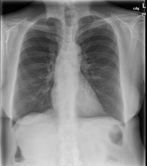
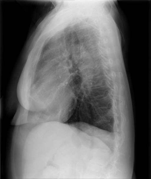
assistant: The chest X-ray images are of good quality, with appropriate patient positioning in both posteroanterior (PA) and lateral views.

Airways appear clear with no signs of hilar adenopathy or enlargement. Breast shadows and bones show no displaced rib fractures or obvious nondisplaced rib fractures, and the remaining bony structures are unremarkable. The cardiomediastinal contour is normal, featuring a typical cardiac silhouette and chamber size, with costophrenic angles being clear, suggesting no pleural effusion. The diaphragm is positioned normally without evidence of free subdiaphragmatic air. Edges reveal no pneumothorax, pleural thickening, or other apical abnormalities. Lung fields are clear bilaterally, with no signs of alveolar air space disease or vascular prominence, and the pulmonary vasculature appears normal. The hilar structures show normal position and vessel formation, with no intrinsic abnormalities noted.

The patient's age and sex are not provided, but the X-ray findings are consistent with a normal examination, devoid of any acute cardiopulmonary or bony pathology.
|
29
|
iu_xray__00000028
|
|
|
user: You are presented with two images, <|img1|> and <|img2|>. Provide a description and your diagnosis.
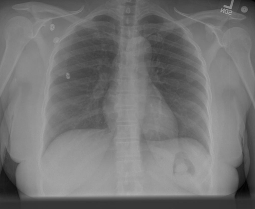
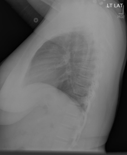
assistant: The provided chest X-ray images include an upright posterior-anterior (PA) view and a left lateral view. The image quality is appropriate with satisfactory contrast and correct patient positioning.

Airways: Trachea is midline with no evidence of any displacement or abnormal opacity in the hilar regions.

Breast shadows and Bones: Breast shadows appear symmetrical. Bones show no signs of rib fractures, lytic lesions, or any bony anomalies.

Cardiomediastinal contour: The heart size is within normal limits, with a normal cardiac silhouette observed. No mediastinal widening is present, and the costophrenic angles are sharp without signs of pleural effusion.

Diaphragm: Both hemidiaphragms are in the normal position, with no subdiaphragmatic air suggesting perforation.

Edges: No evidence of pneumothorax. No pleural thickening, plaques, or fibrosis noted, and extrathoracic tissues appear normal.

Fields: Lung fields are clear with no evidence of alveolar air space disease. There is symmetry between both lungs, with no signs of vascular prominence or other abnormalities.

Hilum: The hilum appears in a normal position with no enlargement. Vascular structures are normal, and there are no masses visible.

Overall, the chest X-rays demonstrate normal findings with clear lungs, normal heart size, and no remarkable abnormalities.
|
30
|
iu_xray__00000029
|
|
|
user: Two images are provided: <|img1|> and <|img2|>. Analyze them and provide your diagnosis.
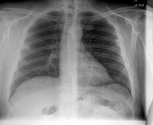
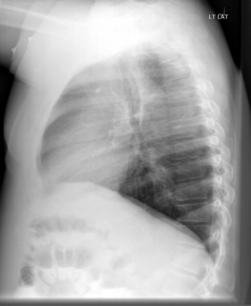
assistant: The chest X-ray images consist of a PA and lateral view. Image quality is adequate with good patient positioning and appropriate contrast.

Airways: The trachea and main bronchi are clear without signs of hilar adenopathy or enlargement.

Breast shadows and bones: No rib fractures or lytic bone lesions are observed. The bones appear intact and well-aligned with no rib crowding.

Cardiomediastinal contour: The cardiac silhouette is normal with no enlargement. The mediastinum and central structures, including clear costophrenic angles, show no signs of pleural effusion.

Diaphragm: Both hemidiaphragms are well-positioned and there is no free air suggesting no abdominal perforation.

Edges: No evidence of pneumothorax. Pleural spaces are clear without thickening, plaques, or fibrosis. Extrathoracic tissues show no abnormalities.

Fields: The lung parenchyma is clear across upper, middle, and lower zones bilaterally, with no asymmetry, alveolar air space disease, or vascular prominence.

Hilum: Normal positioning with no vascular abnormalities. The superior pulmonary vein and inferior pulmonary artery appear normal.

The patient appears to be an adult. No significant findings are noted, and the overall assessment is consistent with a normal chest X-ray.
|
31
|
iu_xray__00000030
|
|
|
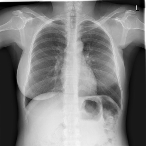
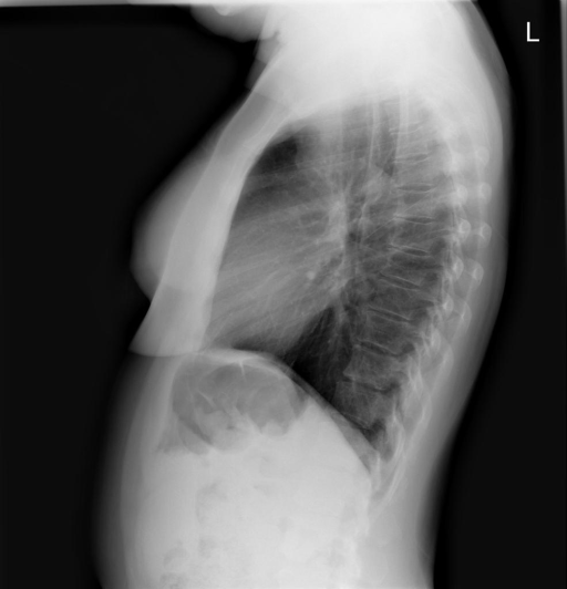
user: Here are two images: <|img1|> and <|img2|>. Explain their contents and share your diagnosis.
assistant: The provided chest X-rays include a posteroanterior (PA) and lateral view, both of good quality with appropriate contrast. The patient positioning appears correct.

The airways are normal, with no evidence of hilar enlargement. The bone structures reveal no rib fractures or lytic lesions; however, left breast postsurgical changes are evident.

The cardiomediastinal silhouette is normal, with the heart and great vessels appropriately sized and shaped. The costophrenic angles are sharp, suggesting no pleural effusions.

Both hemidiaphragms are well-positioned without free air beneath, indicating no signs of abdominal viscus perforation.

The lung fields appear clear in all zones, without signs of consolidation, air space disease, or lymphadenopathy. There is no evidence of pneumothorax, pleural thickening, or fibrosis.

The hila are in normal position, and vessels appear unremarkable.

Demographics: Female (age not provided).

Overall, the X-rays suggest stable postsurgical changes with otherwise normal findings in the chest.
|
32
|
iu_xray__00000031
|
|
|
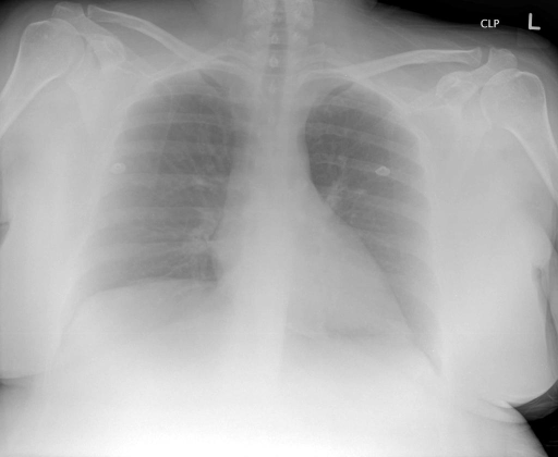
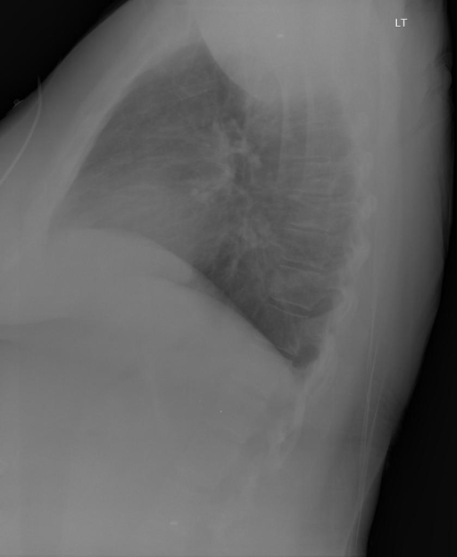
user: Given the two images <|img1|> and <|img2|>, describe their contents and give your diagnosis.
assistant: The chest X-rays include anteroposterior (AP) and lateral views. The image quality is adequate, with appropriate patient positioning.

Airways: Trachea appears midline with no apparent signs of hilar adenopathy or enlargement.

Breast shadows and Bones: The osseous structures, including ribs and vertebrae, show no evidence of fractures or lesions. Bone density appears normal.

Cardiomediastinal contour: The cardiac silhouette is within normal limits. The mediastinum is unremarkable, without widening or abnormal contours. Costophrenic angles are sharp with no pleural effusion noted.

Diaphragm: Both hemidiaphragms are at normal levels, with no free air observed beneath them.

Edges: No pneumothorax is visible. The pleura appear smooth with no thickening or plaques.

Fields: Lung parenchyma is hypoinflated but clear. There is no evidence of focal consolidation, alveolar airspace disease, or increased vascular prominence. Both sides appear symmetrical.

Hilum: Normal vascular structures are observed at the hila, with no displacement or enlargement.

Overall, the X-ray findings suggest clear lungs with normal cardiac and mediastinal structures, though there is noted lung hypoinflation. Further clinical correlation is recommended, especially considering the patient's age and sex if available.
|