| # | key | img1.png | txt |
|---|
1
|
ddti__00000000
|
|
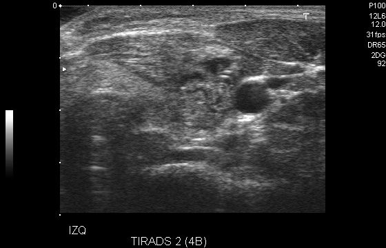
This image identifies the thyroid's position as mostly center, and top-center. The patient is 24, female from Colombia. Observations: microcalcification, well defined smooth margins, tirads 4a. This is a neck ultrasound using a linear transducer, typically in B-mode. It is likely focused on thyroid imaging.

Visible Organs and Structures: The thyroid gland is visible. There is also a nodule present. The gland and surrounding soft tissues are moderately well visualized.

Features of Each Organ/Structure:
- Thyroid Gland:
 - Size: Appears within normal limits for thyroid volume.
 - Shape: Generally regular with slight asymmetry due to the nodule.
 - Texture (echotexture): Primarily homogeneous with a noted hypoechoic nodule.
 - Borders: Well-defined margins of the thyroid.
 - Symmetry: The nodule introduces asymmetry to the affected lobe.

Additional Findings:
- Nodule:
 - Size: Not specified; visually occupies a small portion of the thyroid lobe.
 - Location: Likely within the left thyroid lobe (IZQ suggests "izquierda" or "left").
 - Echotexture: Predominantly hypoechoic.
 - Composition: Solid components, no significant cystic changes.
 - Borders: Fairly smooth, suggesting a low suspicion for malignancy per TIRADS 2 classification.

Gray Scale and Doppler Features:
- Hypoechoic region (nodule) indicates potential benignity given the TIRADS score.
- Doppler studies pending for vascular assessment, though not visible in this image.

Dynamic Features:
- Not applicable as static image provided.

Image Quality and Limitations: Image quality appears fair with slight potential for limitation due to patient positioning or minor soft tissue interference.

Patient Demographic Context: Limited demographic details available; context on patient age and sex would enhance diagnostic interpretation.

Impression/Conclusion: The ultrasound reveals a hypoechoic nodule within the left lobe of the thyroid, classified as TIRADS 2, which is generally considered benign. Follow-up may be warranted based on clinical context and patient history.
|
2
|
ddti__00000001
|
|
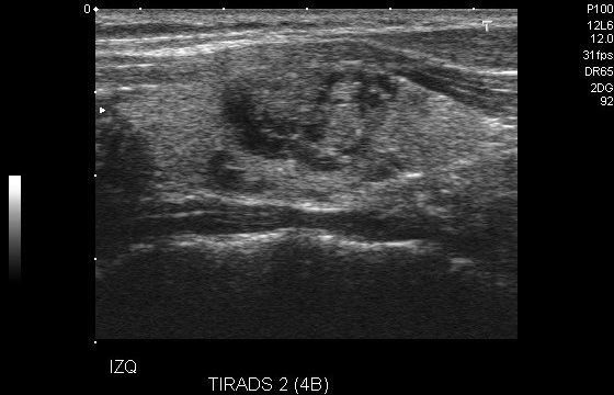
The patient is 24, female from Colombia. Observations: tirads 4a, microcalcification, well defined smooth margins. The thyroid appears to be situated mostly top-center, and center. Thyroid ultrasound using a linear transducer in B-mode. The image primarily displays the thyroid gland.

Visible Organs and Structures: The thyroid gland is well-visualized with surrounding soft tissue structures.

Features of Each Organ/Structure:

- Thyroid Gland:
 - Size: Appears within normal limits; precise measurements are not provided.
 - Shape: Regular, without obvious deformities.
 - Texture (echotexture): Predominantly heterogeneous with areas of isoechoic and hypoechoic regions.
 - Borders: Smooth and well-defined.
 - Symmetry: Only the left lobe is visible; unable to compare bilaterally.

Additional Findings:

- Fluid Presence: No significant fluid collections noted.
- Artifacts: Some posterior acoustic shadowing and enhancement are present but do not impact interpretation.
- Masses/Lesions: A nodule is present, classified as TI-RADS 2, indicating low suspicion for malignancy. It appears hypoechoic with smooth borders.
- Vascular Features: Doppler study not provided, so no vascular features are assessed.

Gray Scale and Doppler Features:

- Hyperechoic: Not prominently noted in the visible thyroid tissue.
- Hypoechoic: Present in the nodule, indicating possible cystic or fibrous content.
- Anechoic: Not observed.

Dynamic Features: Unable to assess due to the static nature of the image.

Image Quality and Limitations: Image quality is good with adequate visualization of the thyroid gland. However, the absence of Doppler limits vascular assessment, and only a portion of the thyroid is visualized.

Patient Demographic Context: Age and sex are not provided.

Impression/Conclusion: The ultrasound reveals a TI-RADS 2 thyroid nodule in the left lobe, suggesting a low likelihood of malignancy. Regular follow-up with ultrasound may be recommended to monitor any changes. Further evaluation with clinical correlation is advised.
|
3
|
ddti__00000002
|
|
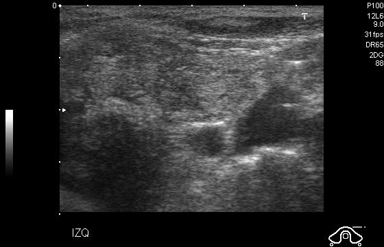
Thyroid placement is reported as mostly center, and top-center. The patient is 74, female from Colombia. Observations: microcalcifications, spiculated margins, solid composition, tirads 4b, hyperechogenicity. This is an abdominal ultrasound using a curvilinear transducer in B-mode. The image displays abdominal anatomical structures, although specific organs are difficult to identify due to the quality or angle of the image.

Visible Organs and Structures:
- The image shows soft tissue structures, but specific organs are poorly visualized.

Features of Each Organ/Structure:
- Due to the limitations in visualization, specific size, shape, and echotexture characteristics of organs are not discernible. Borders are not well-defined, and symmetry cannot be assessed.

Additional Findings:
- Fluid Presence: No distinct fluid collections are visible.
- Artifacts: There appears to be some shadowing and possibly some enhancement in the image.
- Masses/Lesions: No clear masses or lesions can be identified.
- Vascular Features: Doppler analysis is not provided, so vascular flow cannot be assessed.

Gray Scale and Doppler Features:
- Hyperechoic and hypoechoic regions are apparent, but their clinical significance is unclear due to visualization issues.
- Doppler data is not available.

Dynamic Features:
- Due to the static nature of the provided image, organ motion and valvular motion are not observable.

Image Quality and Limitations:
- The image quality is fair, with potential limiting factors including positioning or bowel gas obscuring clear visualization of specific structures.

Patient Demographic Context:
- Patient age and sex are not provided, limiting specific demographic-related interpretation.

Impression/Conclusion:
- This abdominal ultrasound image lacks sufficient clarity to make definitive observations about specific organs or abnormalities. Further imaging with improved visualization may be necessary to draw clinical conclusions.
|
4
|
ddti__00000003
|
|
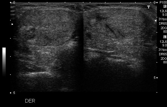
Thyroid placement is reported as top-right, top-left. The patient is 39, female from Colombia. Observations: macrocalcifications, isoechogenicity, well defined margins, predominantly solid composition, tirads 4a. This is an abdominal ultrasound image performed using a linear transducer in B-mode.

Visible Organs and Structures: The image primarily shows soft tissue structures which appear to be parts of the abdomen. Difficult to specify organs without further context, but possibly includes soft tissues like muscles.

Features of Each Organ/Structure:
- Size and Shape: The visible tissues appear regular in shape, but specific measurements or abnormalities cannot be identified from this image alone.
- Texture (echotexture): The echotexture appears mostly homogeneous with regions of varying echogenicity.
- Borders: Borders are moderately defined, though some areas appear slightly indistinct due to the quality of the image.

Additional Findings:
- Fluid Presence: No obvious fluid collections noted in this image.
- Artifacts: Some shadowing is present which may suggest underlying structures or tissues with different densities.
- Masses/Lesions: No discrete masses or lesions are evident in this particular image.
- Vascular Features: Not applicable as Doppler imaging is not used here.

Gray Scale and Doppler Features: The hyperechoic areas could indicate denser tissues, such as tendons or connective tissue, while hypoechoic areas might correspond to muscle or adipose tissue. No Doppler findings are available.

Dynamic Features: Not assessable on this static image.

Image Quality and Limitations: The image quality appears fair; however, the definition is limited, impacting the ability to clearly identify specific anatomical structures. Bowel gas or patient positioning might be factors impacting the clarity.

Patient Demographic Context: Patient age and sex are not provided, limiting contextual interpretation.

Impression/Conclusion: The ultrasound image reveals no obvious abnormalities in the soft tissues visible. However, identification of specific organs or further diagnostic impressions is limited by image quality and lack of Doppler analysis. Further clinical context and additional imaging could aid in a comprehensive assessment.
|
5
|
ddti__00000004
|
|
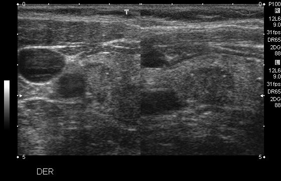
The patient is 40, male from Colombia. Observations: hypoechogenicity, well defined margins, microcalcifications, tirads 5, solid composition. Ultrasound findings indicate the thyroid is mostly center-right, and center. This is a soft tissue ultrasound of the neck using a linear transducer in B-mode.

Visible Organs and Structures: The image shows superficial neck tissues, including skin, subcutaneous tissue, and deeper muscular structures. The thyroid gland and lymph nodes are evident.

Features of Each Organ/Structure:
- Thyroid Gland: Appears to be well-visualized. Size is not directly measurable from the image, but it seems to be within normal size based on comparison with adjacent structures. The texture is homogeneous with hyperechoic presentation. Borders are smooth. No asymmetry noted.
- Lymph Nodes: Several lymph nodes are visible. They are relatively normal in size and appear oval, which is typical. The texture is homogeneous with a slightly hypoechoic center and hyperechoic hilum. Borders are well-defined.

Additional Findings:
- Fluid Presence: No significant fluid collections are observed.
- Artifacts: Mild shadowing behind dense structures, typical in neck ultrasounds.
- Masses/Lesions: No suspicious masses or lesions are observed in this image.
- Vascular Features: Vascular structures are not clearly visible in this image; Doppler imaging would be required for further assessment.

Gray Scale and Doppler Features:
- Hyperechoic Regions: Normal connective tissue and gland outline.
- Hypoechoic Regions: Present in the lymph node centers, indicating normal architecture.

Dynamic Features: Organ motion is not assessed here due to static imaging.

Image Quality and Limitations: The image quality is good; however, minor shadowing is present, likely due to patient positioning or dense tissue.

Patient Demographic Context: Patient age and sex are not provided.

Impression/Conclusion: This ultrasound of the neck shows normal-appearing thyroid tissue and lymph nodes with no abnormalities detected. Given the absence of suspicious findings, the structures seem typical. Further evaluation with Doppler may be useful for assessing vascular flow if clinically indicated.
|
6
|
ddti__00000005
|
|
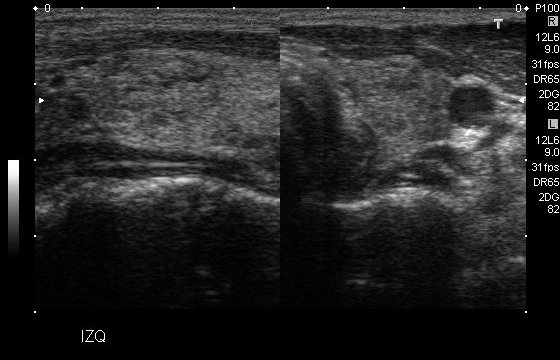
Ultrasound findings indicate the thyroid is top-right, top-center. The patient is 28, female from Colombia. Observations: tirads 4b, microcalcifications, well defined margins, solid composition, isoechogenicity. This is an abdominal ultrasound using a linear transducer in B-mode.

Visible Organs and Structures: The image predominantly shows soft tissues, but specific organs or structures are not clearly labeled or identifiable. It appears to be a cross-sectional view that might include muscle layers and subcutaneous tissue.

Features of Each Organ/Structure:
- Size: No identifiable organs are visible to assess size.
- Shape: Structures visible appear linear and consistent with layered tissue.
- Texture (echotexture): The echotexture appears mixed with areas of hyperechoic and hypoechoic regions suggestive of normal soft tissues.
- Borders: Borders of the visible structures are generally smooth.
- Symmetry: Since there is no clear bilateral structure, symmetry is not applicable.

Additional Findings:
- Fluid Presence: No significant fluid collections are evident in the image.
- Artifacts: Some shadowing and enhancement can be observed, which might affect clarity.
- Masses/Lesions: No distinct masses or lesions are observed.
- Vascular Features: Not applicable as vessels are not clearly visible.

Gray Scale and Doppler Features: Hyperechoic regions represent denser tissue interfaces, while hypoechoic regions indicate areas of less density. No Doppler features are shown in this still image.

Dynamic Features: Dynamic motion features are not assessable in this static image.

Image Quality and Limitations: The image quality appears fair, but it may be limited by the display of a narrow field of view. The lack of identifiable landmarks or labeling restricts specific interpretation.

Patient Demographic Context: Information on patient age and sex is not provided.

Impression/Conclusion: The ultrasound image shows normal mixed echotexture of soft tissues in the imaged area without clear pathology. The lack of visible pathology or additional patient context limits diagnostic consideration. A more comprehensive evaluation with labeled anatomy or live imaging could enhance interpretation.
|
7
|
ddti__00000006
|
|
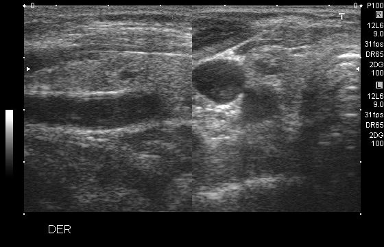
The thyroid gland's location is primarily top-center, top-left. The patient is 60, female from Colombia. Observations: well defined margins, isoechogenicity, spongiform composition, microcalcifications, tirads 4a. The image appears to be from a musculoskeletal ultrasound using a linear transducer in B-mode.

Visible Organs and Structures: The structures visible seem to be soft tissue, potentially part of a limb, with visible muscle and possibly a joint or tendon.

Features of Each Organ/Structure:
- The muscle tissue appears well-visualized with a homogeneous echotexture.
- A round anechoic area, possibly a cyst or small fluid collection, is visible.
- Borders of the anechoic area look smooth and well-defined.
- No obvious asymmetry noted in the structures.

Additional Findings:
- There is an anechoic region suggesting a fluid-filled cystic area.
- No noteworthy artifacts such as shadowing or enhancement present.
- No masses or lesions with irregular borders identified.

Gray Scale and Doppler Features: Hyperechoic and hypoechoic regions are indicative of normal muscle and connective tissue contrast. Doppler features are not available in this image.

Dynamic Features: Not assessable in this static image.

Image Quality and Limitations: The image quality appears fair, but interpretation may be limited due to lack of Doppler data or motion assessment features.

Patient Demographic Context: Not provided; would be useful for thorough interpretation.

Impression/Conclusion: The ultrasound shows a normal appearance of the muscle tissue with a possible small cyst or fluid collection. No concerning lesions or abnormalities noted. Further clinical correlation and additional imaging may be warranted to characterize the anechoic area if symptomatic.
|
8
|
ddti__00000007
|
|
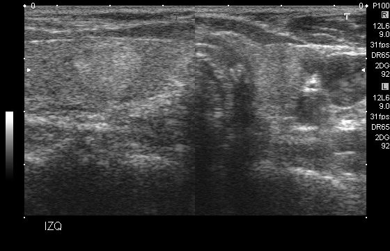
The patient is 82, female from Colombia. Observations: hyperechogenicity, tirads 4a, solid composition, well defined margins. The thyroid gland's location is primarily top-right. This is an ultrasound image, likely soft tissue. The type of transducer appears to be linear, typically used for superficial structures. The mode is B-mode, which provides a grayscale image.

Visible Organs and Structures: The image primarily shows soft tissue structures. Specific organs are not distinctly visualized, suggesting this could be an image of a body region like the musculoskeletal area.

Features of Each Organ/Structure:
- Size: Not applicable due to lack of distinct organs.
- Shape: Not specified due to lack of identifiable structures.
- Texture (echotexture): The overall echotexture appears heterogeneous with varying shades of gray.
- Borders: Some areas suggest ill-defined borders due to overlapping tissue densities.
- Symmetry: Not applicable as it is a single-plane image without bilateral comparison.

Additional Findings:
- Fluid Presence: No obvious anechoic or hypoechoic regions indicating fluid collections.
- Artifacts: Some shadowing is present, typical in soft tissue imaging.
- Masses/Lesions: No distinct masses or lesions observed.
- Vascular Features: Not assessed as Doppler is not activated.

Gray Scale and Doppler Features: The presence of hypoechoic regions could suggest denser tissue areas, but no Doppler findings are available in the static image.

Dynamic Features: Dynamic assessment is not possible with the static image provided.

Image Quality and Limitations: Image quality appears fair. The clarity might be affected by patient positioning or interference from surrounding tissues.

Patient Demographic Context: Not provided. Typically, age and sex could influence interpretation, especially if the ultrasound targets specific organs.

Impression/Conclusion: This ultrasound image primarily captures soft tissue with no distinct organs visualized. The heterogeneous echotexture does not suggest any specific pathology without further context. A follow-up with more detailed imaging or clinical correlation may be needed.
|
9
|
ddti__00000008
|
|
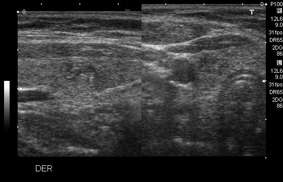
The patient is 41, female from Colombia. Observations: tirads 4a, spongiform composition, isoechogenicity, well defined margins, microcalcifications. The thyroid gland's location is primarily center-right. The image appears to be from a musculoskeletal ultrasound, likely using a linear transducer in B-mode. This is often used for detailed imaging of superficial structures such as tendons, ligaments, and skin.

Visible Organs and Structures: The image shows the dermis and subcutaneous tissue. No deeper structures are identifiable, likely due to the positioning and focus of the ultrasound.

Features of Each Organ/Structure:
- Dermis and Subcutaneous Tissue:
 - Size: Normal size for the visible layers.
 - Shape: Regular structure with no apparent deformities.
 - Texture (echotexture): The dermis displays a homogeneous pattern, while subcutaneous tissue appears slightly heterogeneous due to natural fat lobules.
 - Borders: The border between the dermis and subcutaneous tissue is smooth and well-defined.
 - Symmetry: Not applicable as it’s focusing on a single area.

Additional Findings:
- Fluid Presence: No obvious fluid collections visible in the image.
- Artifacts: Minimal artifacts, with slight reverberation common in soft tissue imaging.
- Masses/Lesions: None detected.
- Vascular Features: Not assessed; Doppler not utilized in this image.

Gray Scale and Doppler Features:
- No significant hyperechoic or hypoechoic regions indicating abnormal findings in the visible area.

Dynamic Features:
- Not applicable for this static ultrasound image as motion evaluation is not possible.

Image Quality and Limitations: The image quality is good, with clear visualization of superficial tissues. Limitations include inability to visualize deeper structures, which could be due to the focus on superficial layers and potential presence of overlying artifacts.

Patient Demographic Context: The image lacks context about patient age and sex, which would help tailor assessments, particularly in musculoskeletal evaluations.

Impression/Conclusion: The ultrasound shows normal appearing dermal and subcutaneous structures without any visible pathologies such as fluid collections or masses. If clinical symptoms or concerns warrant further investigation, additional imaging or modalities might be considered.
|
10
|
ddti__00000009
|
|
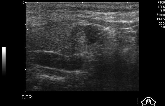
The current scan suggests the thyroid is positioned mostly top-center, and center. The patient is 73, female from Colombia. Observations: tirads 4a, microcalcifications, predominantly cystic composition, well defined margins, isoechogenicity. This is an abdominal ultrasound using a linear transducer in B-mode.

Visible Organs and Structures: The image primarily shows soft tissue structures, possibly indicating a region in the abdominal area. Exact organs are not specified, but it appears to include layers of muscle or connective tissue.

Features of Each Organ/Structure:
- Size: Cannot determine specific organ size as individual organs are not identifiable.
- Shape: The visible structures appear linear, typical of muscular or fibrous tissue.
- Texture (echotexture): There is a combination of hyperechoic (brighter) and hypoechoic (darker) areas, suggesting mixed echotexture.
- Borders: The borders of the visible structures are fairly smooth but not distinctly defined.
- Symmetry: Bilateral structures cannot be assessed from this image.

Additional Findings:
- Fluid Presence: No obvious fluid collections noted.
- Artifacts: Minimal shadowing and acoustic enhancement are observed, likely due to the soft tissue composition.
- Masses/Lesions: No distinct masses or lesions identified.
- Vascular Features: Not assessable in this image as Doppler mode is not utilized.

Gray Scale and Doppler Features:
- The hyperechoic areas may represent dense tissues such as fascia, while hypoechoic areas may indicate muscle layers.
- Doppler assessment is not applicable here.

Dynamic Features: No apparent motion features are visible, as this is a static image.

Image Quality and Limitations: Image quality is fair, but tissue delineation is limited. No clear visualization of abdominal organs due to the potential presence of overlying tissue and lack of anatomical landmarks.

Patient Demographic Context: Not provided.

Impression/Conclusion: This ultrasound image shows mixed echotexture of soft tissue, potentially within the abdominal region. No specific abnormalities are appreciable. For a thorough assessment, correlative imaging with identified landmarks or additional clinical information may be recommended.
|
11
|
ddti__00000010
|
|
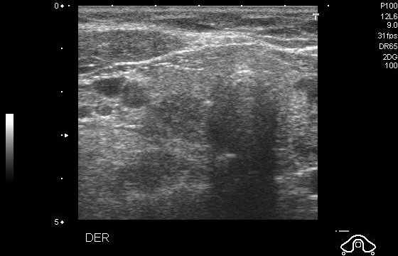
The patient is 50, female from Colombia. Observations: tirads 4b, well defined margins, hypoechogenicity, predominantly cystic composition, microcalcifications. The position of the thyroid is described as center. The image is likely from a breast ultrasound, utilizing a linear transducer in B-mode.

Visible Organs and Structures: The primary structure visible is breast tissue, appearing well-visualized.

Features of Each Organ/Structure:
- Breast Tissue:
 - Size: Specific measurements are not provided, but the tissue appears to be of normal size for ultrasound imaging.
 - Shape: The breast tissue has a regular contour with no obvious deformities.
 - Texture (echotexture): There is a heterogeneous echotexture with areas of mixed echogenicity.
 - Borders: The tissue borders seem smooth but may be obscured in some regions due to artifacts.
 - Symmetry: Not applicable, as this is a unilateral image.

Additional Findings:
- Fluid Presence: No significant fluid collections are noted in the imaged area.
- Artifacts: There is posterior acoustic shadowing visible, common in breast ultrasounds.
- Masses/Lesions: No distinct masses or lesions are observed in this image.
- Vascular Features: Not noted in this image, Doppler imaging was not applied.

Gray Scale and Doppler Features:
- Hyperechoic and hypoechoic regions are present but are not indicative of any specific condition based on this single image.

Dynamic Features: Due to the static nature of the image, dynamic features such as tissue motion are not assessable.

Image Quality and Limitations: The image quality appears to be good, though obscured areas by shadowing may impact full interpretation.

Patient Demographic Context: The patient's age and sex are not provided, but typically breast ultrasounds are performed on females for evaluation of palpable masses or routine screening.

Impression/Conclusion: The ultrasound image shows breast tissue with a heterogeneous echotexture and some artifacts but no overt lesions identifiable. Further clinical context or additional imaging might be needed for comprehensive evaluation.
|
12
|
ddti__00000011
|
|
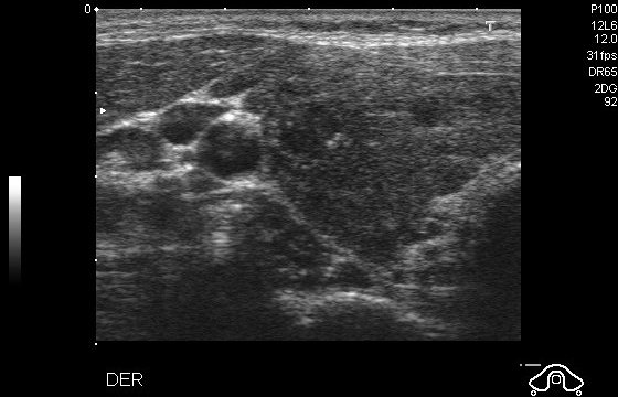
The patient is 30, female from Colombia. Observations: microcalcifications, marked hypoechogenicity, solid composition, spiculated margins, tirads 5. The thyroid is predominantly mostly top-center, and center. This is a soft tissue ultrasound, probably using a linear transducer in B-mode for detailed structural visualization.

Visible Organs and Structures: The image shows subcutaneous tissue and possibly some nodular formations. The location is likely superficial given the use of a linear transducer.

Features of Each Organ/Structure:
- Nodular Structures:
 - Size: Two nodules are apparent; size measurements aren’t provided, but they appear relatively small.
 - Shape: Roughly round nodules.
 - Texture (echotexture): Predominantly hypoechoic with potential anechoic areas, suggesting cystic components.
 - Borders: Well-defined.

Additional Findings:
- Fluid Presence: No distinct fluid collection noted.
- Artifacts: Acoustic enhancement might be present beneath the nodules.
- Masses/Lesions: The nodules could suggest cysts or lymph nodes; however, further clinical correlation is needed.
- Vascular Features: Doppler was not used in this image, so no flow patterns can be described.

Gray Scale and Doppler Features: The hypoechoic areas suggest a possible fluid component or different tissue type compared to surrounding areas. Doppler findings are not applicable here.

Dynamic Features: Not applicable as this is a static image.

Image Quality and Limitations: The image quality appears fair, with some graininess; interpretation might be limited by patient positioning or superficial nature limiting deeper tissue visualization.

Patient Demographic Context: Specific patient age and sex are not provided, which would help in contextualizing findings.

Impression/Conclusion: The ultrasound shows well-defined hypoechoic nodules in the subcutaneous tissue, suggestive of cystic lesions or lymph nodes. Further evaluation with clinical correlation and potentially Doppler imaging could provide more insights.
|
13
|
ddti__00000012
|
|
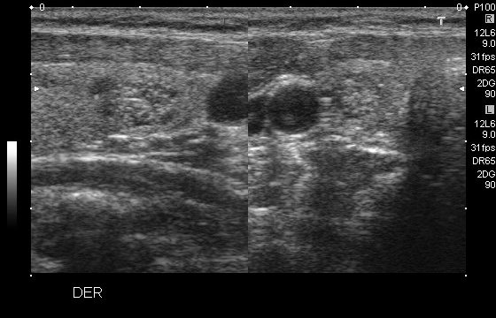
The patient is 56, female from Colombia. Observations: well defined margins, microcalcifications, hyperechogenicity, predominantly solid composition, tirads 4a. The current scan suggests the thyroid is positioned top-right. This appears to be a soft tissue ultrasound, likely using a linear transducer in B-mode.

Visible Organs and Structures: The image shows subcutaneous tissue and possibly part of a superficial structure, such as a vein or cyst. Limited deeper structures are visible due to the field of view.

Features of Each Organ/Structure:
- Visible Structure: Likely a cystic area near the center.
 - Size: Unable to measure accurately; appears small.
 - Shape: Roughly round.
 - Texture (echotexture): Anechoic, suggesting a fluid-filled nature.
 - Borders: Well-defined.
 - Symmetry: Not applicable as only one structure is visible clearly.

Additional Findings:
- Fluid Presence: Anechoic area suggesting a fluid-filled cyst.
- Artifacts: Minimal artifacts; good acoustic shadowing indicative of fluid.
- Masses/Lesions: The anechoic structure may suggest a benign cyst.
- Vascular Features: Not assessed due to lack of Doppler imaging in this frame.

Gray Scale and Doppler Features:
- Hyperechoic and hypoechoic areas are consistent with typical soft tissue and fluid interfaces.
- Doppler findings absent due to the mode used.

Dynamic Features: Not applicable in this single-frame image.

Image Quality and Limitations:
The image quality appears fair. Potential limitations include limited field depth and absence of Doppler, which restricts vascular assessment.

Patient Demographic Context: Not provided.

Impression/Conclusion: The ultrasound image shows a possibly benign cystic area within subcutaneous tissue. Further correlation with clinical findings and possibly Doppler ultrasound for vascular assessment would be needed for comprehensive evaluation.
|
14
|
ddti__00000013
|
|
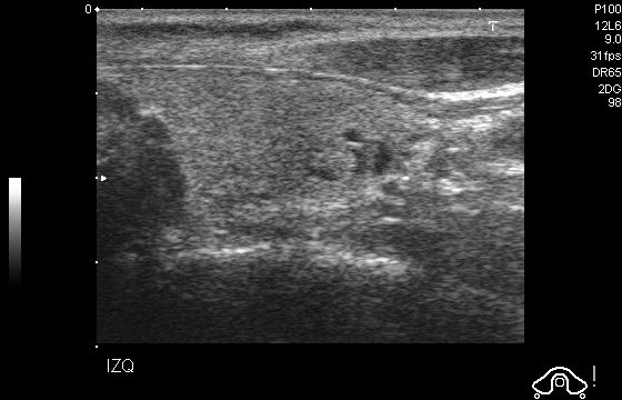
The patient is 34 from Colombia. Observations: spongiform composition, isoechogenicity, microcalcifications, well defined margins, tirads 4a. Observed thyroid position: center. This is an abdominal ultrasound using a curvilinear transducer in B-mode.

Visible Organs and Structures: The image appears to show the liver. The organ is well-visualized, though the image quality could be better for more detailed assessment.

Features of Each Organ/Structure:
- Liver:
 - Size: Cannot be measured accurately in this image.
 - Shape: Regular.
 - Texture (echotexture): Appears homogeneous.
 - Borders: Smooth and well-defined.
 - Symmetry: Not applicable to the liver itself, but needs comparison with the contralateral lobe if possible.

Additional Findings:
- Fluid Presence: No fluid collections are noted in this image.
- Artifacts: Some artifacts might be present, such as shadowing or noise due to image quality.
- Masses/Lesions: No masses or lesions are visible in this image.
- Vascular Features: Not assessed due to absence of Doppler mode.

Gray Scale and Doppler Features: There's distribution of hyperechoic and hypoechoic areas indicating normal liver tissue contrast. Doppler features are not present as the image does not include Doppler mode.

Dynamic Features: Not applicable as this is a static image.

Image Quality and Limitations: The image quality is fair; limited resolution may impact the assessment of subtle pathologies.

Patient Demographic Context: Not provided.

Impression/Conclusion: Liver appears to be normal in size and echotexture. No obvious abnormalities such as masses or lesions are visible. Image quality is fair, potentially limiting the detection of smaller pathologies. Further evaluation with higher quality or Doppler imaging is recommended if clinical symptoms suggest liver pathology.
|
15
|
ddti__00000014
|
|
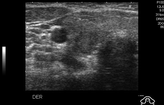
Observed thyroid position: center. The patient is 36, female from Colombia. Observations: microcalcifications, solid composition, ill defined margins, hypoechogenicity, tirads 4c. This image appears to be from a soft tissue ultrasound, most likely using a linear transducer with B-mode imaging.

Visible Organs and Structures: The image depicts subcutaneous tissue and possibly part of a small organ or gland. It seems to lack distinct major organs, indicating a focus on superficial or peripheral structures.

Features of Each Organ/Structure:
- Visible structure appears as a circular or oval shape, possibly a cyst or lymph node.
- Size: Small, though exact measurements aren't provided.
- Shape: Regular and round.
- Texture (echotexture): Hypoechoic center with potentially hyperechoic peripheral border.
- Borders: Clearly defined.
- Symmetry: Not applicable due to singular structure.

Additional Findings:
- Fluid Presence: No obvious fluid collections detected within the observed area.
- Artifacts: A potential posterior acoustic enhancement could suggest cystic nature.
- Masses/Lesions: The hypoechoic round structure may indicate a mass, requiring further evaluation.
- Vascular Features: Not assessable due to lack of Doppler imaging.

Gray Scale and Doppler Features:
- The hypoechoic nature of the round structure may indicate a cyst, though further confirmation with clinical correlation is necessary.

Dynamic Features: Not assessable in this static image.

Image Quality and Limitations: Image quality is good but limited by absence of additional context or structures for broader evaluation. Position and surrounding tissue visualization are adequate.

Patient Demographic Context: Information on patient age and sex is not provided, which could aid in diagnosis.

Impression/Conclusion: The ultrasound shows a well-defined, hypoechoic, likely superficial structure that could be a cyst or lymph node. Further clinical correlation and potentially additional imaging or Doppler study could clarify the diagnosis.
|
16
|
ddti__00000015
|
|
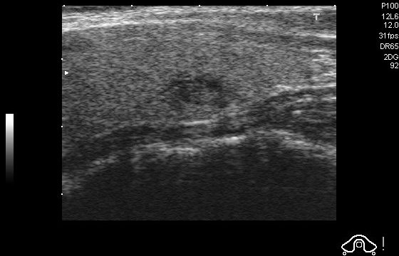
The patient is 27 from Colombia. Observations: isoechogenicity, tirads 4a, well defined margins, spongiform composition. This image identifies the thyroid's position as mostly center, and top-center. This is a musculoskeletal ultrasound, likely using a linear transducer in B-mode, ideal for examining superficial structures.

Visible Organs and Structures: The image shows a portion of soft tissue, possibly muscle or tendon, though detailed identification is limited due to the image's quality.

Features of Each Organ/Structure:
- Size: The structures appear within normal range, though precise measurements are not possible.
- Shape: The soft tissues depicted seem regular, without notable deformities or asymmetry.
- Texture (echotexture): The echotexture is heterogeneous, with a mix of hyperechoic and hypoechoic areas common in musculoskeletal scans.
- Borders: Borders of structures are poorly defined, which may be due to resolution and the absence of clear delineation.

Additional Findings:
- Fluid Presence: No obvious fluid collections are visible in this image.
- Artifacts: There are possible artifacts present, including some shadowing and enhancement.
- Masses/Lesions: There is no clear evidence of masses or lesions within this image.
- Vascular Features: Not applicable in this image due to its nature and mode.

Gray Scale and Doppler Features: The hyperechoic regions may suggest fibrous tissue content, while hypoechoic areas could indicate potential edema or fat. No Doppler findings are available or applicable.

Dynamic Features: As this is a static image, no dynamic features such as organ motion can be assessed.

Image Quality and Limitations: The image quality seems fair but is limited by resolution and potential artifacts, making precise interpretation challenging. There may be influence from overlying structures or patient positioning.

Patient Demographic Context: Not provided.

Impression/Conclusion: This ultrasound image of the musculoskeletal region shows normal echotexture without evidence of abnormalities such as significant fluid collections or lesions. The heterogeneity is typical of musculoskeletal tissues. Further dynamic assessment and clinical correlation are advised for comprehensive evaluation.
|
17
|
ddti__00000016
|
|
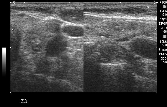
The patient is 76, female from Colombia. Observations: hypoechogenicity, tirads 4c, microcalcifications, solid composition, well defined margins. The thyroid appears to be situated mostly top-right, and top-left. This appears to be a transverse breast ultrasound likely using a linear transducer in B-mode.

Visible Organs and Structures: The primary structure in the image is breast tissue, displaying variable echotexture. There are also indications of ducts or cystic areas.

Features of Each Organ/Structure:
- Size: The structures lack distinct measurements in the provided image. Important structures appear moderately sized.
- Shape: The prominent lesions or structures demonstrate mostly oval and regular shapes.
- Texture (echotexture): The tissue exhibits a heterogeneous echotexture with both hypoechoic and hyperechoic areas.
- Borders: Some areas show well-defined, smooth borders, while others are less distinct.
- Symmetry: As this is a single image frame, symmetry cannot be evaluated.

Additional Findings:
- Fluid Presence: Potential fluid-filled cystic areas are visible, notably anechoic and well-circumscribed.
- Artifacts: No significant acoustic shadowing or enhancement artifacts noted.
- Masses/Lesions: Presence of hypoechoic masses with oval shape, possibly indicative of benign lesions such as fibroadenomas.
- Vascular Features: Not applicable as Doppler mode is not evident.

Gray Scale and Doppler Features:
- Hypoechoic regions likely correspond to solid masses or fluid collections.
- Anechoic regions suggest simple cysts. Doppler features are not evident.

Dynamic Features:
- The static nature of the image means dynamic features cannot be assessed.

Image Quality and Limitations: The image quality appears fair, though resolution and contrast may limit fine detail interpretation. Factors such as patient positioning or potential compression are not impacting this image specifically.

Patient Demographic Context: Not provided, but age and sex (typically female) would be relevant.

Impression/Conclusion: The ultrasound reveals heterogeneous breast tissue with well-circumscribed hypoechoic lesions, likely benign cysts or fibroadenomas. No suspicious features such as irregular borders or posterior acoustic shadowing are apparent in this single image frame. Further evaluation with additional views and clinical correlation is recommended to clarify these findings.
|
18
|
ddti__00000017
|
|
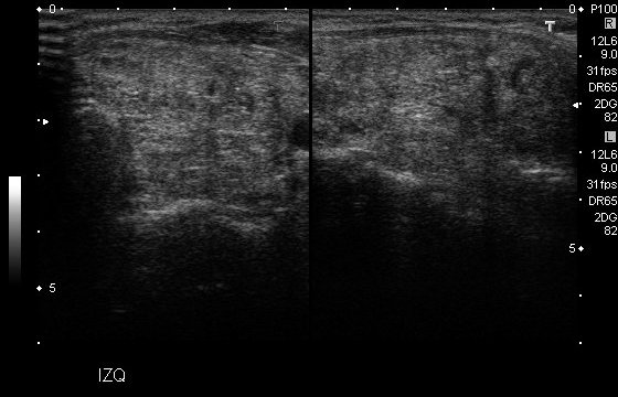
The patient is 64, female from Colombia. Observations: solid composition, tirads 4b, isoechogenicity, microlobulated margins, microcalcifications. Ultrasound findings indicate the thyroid is top-right. This is a breast ultrasound, likely using a linear transducer in B-mode, as these are standard for breast imaging.

Visible Organs and Structures: The primary structure visible is breast tissue, with subcutaneous fat and glandular tissue.

Features of Each Organ/Structure:
- Breast Tissue:
 - Size: Normal; specific size measurements are not displayed.
 - Shape: Regular, typical for breast architecture.
 - Texture (echotexture): Mixed echotexture with both hyperechoic and hypoechoic areas representing normal glandular and fatty tissue.
 - Borders: Smooth, with no significant irregularities noted.
 - Symmetry: This is an isolated view, so comparison with the contralateral side is not possible.

Additional Findings:
- Fluid Presence: No significant fluid collections observed.
- Artifacts: Minimal shadowing, which is typical in heterogeneous tissue.
- Masses/Lesions: From the image provided, no distinct masses or lesions are evident.
- Vascular Features: Not applicable as this is a non-Doppler image.

Gray Scale and Doppler Features:
- Clinical significance of hyperechoic and hypoechoic regions is consistent with normal variation in breast tissue composition.
- Doppler examination not applicable; no flow patterns or waveforms are assessed here.

Dynamic Features:
- Not applicable in static breast ultrasound images.

Image Quality and Limitations: The image quality appears fair, with adequate visualization of breast tissue. Potential limitations include absence of the entire breast and axillary views, which are crucial for comprehensive evaluation. Positioning seems adequate.

Patient Demographic Context: Patient age and sex are not provided but would typically be female for a breast ultrasound. Age is crucial for assessing normalcy of tissue patterns.

Impression/Conclusion: The ultrasound presents normal mixed echotexture of breast tissue with no visible masses or lesions. Given the limitations of the single image, further clinical correlation and possible additional imaging may be warranted to rule out focal findings.
|
19
|
ddti__00000018
|
|
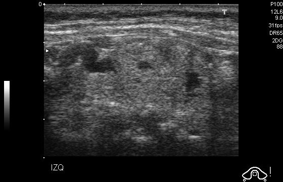
Observed thyroid position: top-center, center-right. The patient is 60, female from Colombia. Observations: microcalcifications, spongiform composition, tirads 4a, well defined margins, isoechogenicity. This is an abdominal ultrasound using a curvilinear transducer in B-mode.

Visible Organs and Structures: The image shows part of the abdominal region. Specific organs are not well-visualized due to image quality.

Features of Each Organ/Structure: Due to the grainy quality of the image, detailed assessment of organs for size, shape, texture, or borders is limited.

Additional Findings:
- Fluid Presence: No distinct fluid collections are visible.
- Artifacts: Graininess suggests possible interference from bowel gas or suboptimal positioning.
- Masses/Lesions: No clear masses or lesions are visible.
- Vascular Features: No Doppler features can be analyzed due to the mode used.

Gray Scale and Doppler Features: The image primarily shows a homogenous echotexture without specific focal hyperechoic, hypoechoic, or anechoic areas identified within the scanned section.

Dynamic Features: Due to the static nature of the image, dynamic features such as organ motion cannot be assessed.

Image Quality and Limitations: Image quality is poor, likely affected by bowel gas or insufficient transducer pressure, limiting detailed evaluation.

Patient Demographic Context: The ultrasound does not provide specific information on patient age or sex.

Impression/Conclusion: The ultrasound image lacks sufficient clarity for a comprehensive evaluation of abdominal structures. No significant findings are observed; further imaging with optimized settings is recommended to improve diagnostic accuracy.
|
20
|
ddti__00000019
|
|
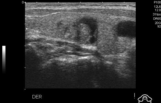
The patient is 45, female from Colombia. Observations: isoechogenicity, microcalcifications, tirads 4a, predominantly solid composition, well defined margins. The current scan suggests the thyroid is positioned mostly top-center, and center. The image appears to be a soft tissue ultrasound, likely using a linear transducer in B-mode.

Visible Organs and Structures: The image primarily shows soft tissue structures. There may be muscles and subcutaneous tissue visualized.

Features of Each Organ/Structure:
- Size: Structures appear within normal size range, though specific measurements are not provided.
- Shape: The visible structures have a regular appearance.
- Texture (echotexture): The echotexture is relatively homogeneous with some hypoechoic and hyperechoic areas.
- Borders: Borders of the structures appear mostly smooth but slightly irregular in areas.
- Symmetry: Symmetry is not directly assessed due to the nature of the image.

Additional Findings:
- Fluid Presence: No evident fluid collections are visible in this image.
- Artifacts: Some shadowing is noted, possibly due to underlying bony structures or dense tissue.
- Masses/Lesions: No distinct masses or lesions are immediately visible.
- Vascular Features: No Doppler mode applied; therefore, no vascular features are described.

Gray Scale and Doppler Features:
- Hyperechoic regions could represent connective tissue or interfaces between different types of tissue.
- Hypoechoic areas may represent muscle or fat tissue; no anechoic areas suggesting fluid are present.

Dynamic Features: Not applicable as this is a static image.

Image Quality and Limitations:
- Image quality appears fair, though lateral resolution could be limited. Details may be impacted by the depth setting and gain.

Patient Demographic Context: No information on patient age or sex is provided in the image or description.

Impression/Conclusion: The soft tissue structures appear normal, with no clear evidence of abnormal lesions or fluid collections. Further clinical correlation and possibly dynamic ultrasound might be needed for additional assessment.
|
21
|
ddti__00000020
|
|
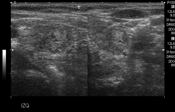
Observed thyroid position: center-left. The patient is 77, female from Colombia. Observations: macrolobulated margins, microcalcifications, solid composition, hyperechogenicity, tirads 4b. This image appears to be a musculoskeletal ultrasound, likely using a high-frequency linear transducer in B-mode.

Visible Organs and Structures: The image shows superficial soft tissue structures. It seems to depict regions that could include muscle or possibly a glandular area, given the texture and layering visible.

Features of Each Organ/Structure:
- Size: Specific measurements are not visible due to the lack of scale.
- Shape: The structures seem to have a relatively regular shape, without obvious deformities.
- Texture (echotexture): The region displays mixed echotexture with both hyperechoic and hypoechoic areas, indicating varied tissue densities.
- Borders: The borders of the observable structures are somewhat irregular, but generally well-defined.
- Symmetry: Not applicable due to a single-side view.

Additional Findings:
- Fluid Presence: A hypoechoic area is present, possibly indicating a fluid collection or cystic structure.
- Artifacts: There is slight acoustic shadowing at deeper levels, common in dense tissues.
- Masses/Lesions: No distinct masses or lesions clearly visible at this scale and quality.
- Vascular Features: No Doppler information available to assess vascular flow.

Gray Scale and Doppler Features: Hyperechoic regions suggest fibrous or calcified tissue, while hypoechoic regions might indicate fluid or less dense tissue. There's no Doppler data on flow patterns or velocities.

Dynamic Features: No motion features are observable in this static image.

Image Quality and Limitations: The image quality is fair, but the resolution limits detailed characterization. Depth and positioning restrict the view of deeper structures.

Patient Demographic Context: Not available in the image.

Impression/Conclusion: The ultrasound shows superficial tissues with varying echogenicity, suggesting possible normal soft tissue anatomy or minor pathology such as a small fluid collection. There is no clear evidence of significant abnormality, but the limitations of the image quality and lack of clinical information should be considered. Further assessment or alternative imaging might be warranted if there's clinical suspicion of pathology.
|
22
|
ddti__00000021
|
|
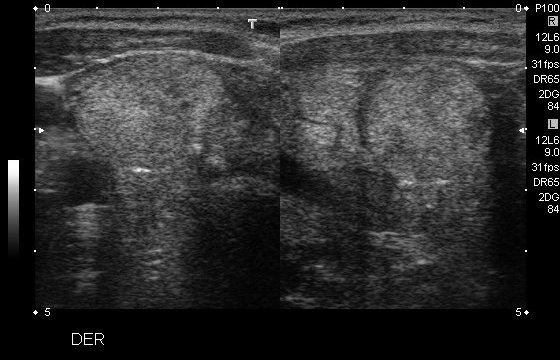
Ultrasound findings indicate the thyroid is . The patient is 80, female from Colombia. Observations: isoechogenicity, solid composition, tirads 4b, well defined margins, microcalcifications. Musculoskeletal ultrasound using a linear transducer in B-mode.

Visible Organs and Structures: The ultrasound image depicts a section of the right shoulder. Visible structures include the deltoid muscle and surrounding soft tissue. The visibility of deeper structures such as the rotator cuff tendons is limited.

Features of Each Organ/Structure:
- Deltoid Muscle:
 - Size: Appears normal; no measurements provided.
 - Shape: Regular without obvious deformities.
 - Texture (echotexture): Homogeneous with normal, consistent echogenicity.
 - Borders: Smooth and well-defined.
 - Symmetry: Unable to assess symmetry as the left shoulder is not imaged.

Additional Findings:
- Fluid Presence: No evidence of fluid collections observed in the imaged region.
- Artifacts: Minor shadowing noted, likely due to patient positioning or scanning angle.
- Masses/Lesions: No masses or lesions are evident.
- Vascular Features: Doppler mode not applied; no assessment of vascular flow.

Gray Scale and Doppler Features:
- Echogenicity of examined structures appears within normal range. No Doppler evaluation provided.

Dynamic Features: No dynamic features such as muscle contraction or joint motion assessable from this static image.

Image Quality and Limitations: Image quality is adequate for assessing superficial structures but limited for deeper anatomical features due to the absence of Doppler and shadowing artifacts. Positioning may affect visualization of deeper structures.

Patient Demographic Context: No patient age or sex information provided. Assuming a general adult demographic unless specified otherwise.

Impression/Conclusion: The image shows a normal-appearing deltoid muscle with no visible abnormalities in the superficial soft tissues of the right shoulder. The assessment of deeper structures such as the rotator cuff is limited due to the lack of depth in imaging and shadowing. Further evaluation with better positioning or imaging modalities like MRI may be considered if deeper assessment is needed.
|
23
|
ddti__00000022
|
|
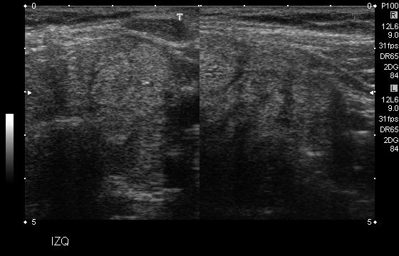
The patient is 80, female from Colombia. Observations: isoechogenicity, tirads 4b, microcalcifications, solid composition, well defined margins. Ultrasound findings indicate the thyroid is . This is a breast ultrasound using a linear transducer in B-mode.

Visible Organs and Structures: The image shows breast tissue. Detailed anatomical structures are not specifically labeled, which is common in general breast ultrasounds, but the overall breast parenchyma is the focus.

Features of Each Organ/Structure:
- Size: Not explicitly measurable from a single static image, but the breast tissue appears to occupy the whole frame.
- Shape: The tissue seen does not display significant irregularities within the provided image frames.
- Texture (echotexture): The breast parenchyma exhibits a heterogeneous echotexture, with alternating hyperechoic and hypoechoic regions indicative of normal glandular and fatty tissue variance.
- Borders: No distinct borders of masses or lesions are visible.
- Symmetry: The image is of a single breast, so symmetry assessment is not applicable.

Additional Findings:
- Fluid Presence: No obvious anechoic fluid collections indicating cysts.
- Artifacts: The image appears to have some shadowing or enhancement, which can be common in breast tissue due to glandular structure.
- Masses/Lesions: No definitive masses or lesions are noted.
- Vascular Features: Not assessable as Doppler mode is not utilized here.

Gray Scale and Doppler Features:
- Hyperechoic regions likely represent fibroglandular tissue, whereas hypoechoic regions could represent adipose tissue. There is no Doppler information present.

Dynamic Features:
- This static image does not allow assessment of dynamic features like motion.

Image Quality and Limitations: The image quality appears fair. Limitations include the static nature of the image and potential artifacts which could obscure small lesions or abnormalities.

Patient Demographic Context: Patient age and sex are not provided, although the examination of breast tissue implies a female patient.

Impression/Conclusion: This ultrasound image of the breast shows a normal heterogeneous echotexture typical of breast parenchyma with no evident masses, fluid collections, or distinct lesions. Further dynamic study or Doppler assessment might be useful if indicated by clinical presentation or further suspicion of pathology.
|
24
|
ddti__00000023
|
|
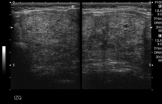
Observed thyroid position: top-left. The patient is 47, female from Colombia. Observations: tirads 4c, solid composition, hyperechogenicity, microcalcifications, ill defined margins. This is an abdominal ultrasound using a linear transducer in B-mode.

Visible Organs and Structures: The image primarily shows soft tissue structures. Specific identification is limited due to the grayscale image and lack of clear landmarks typically seen in labeled ultrasound images.

Features of Each Organ/Structure:

- Size: Not applicable; specific structures are not labeled.
- Shape: Indeterminate due to lack of identifiable structures.
- Texture (echotexture): The visible tissue appears heterogeneous, with a mix of hyperechoic and hypoechoic regions.
- Borders: The borders of the visible tissue area are relatively smooth but not sharply defined.
- Symmetry: Not applicable in this view.

Additional Findings:

- Fluid Presence: No clear anechoic (fluid-filled) areas are noted.
- Artifacts: Some shadowing and signal dropout are present, which is typical in soft tissue ultrasound.
- Masses/Lesions: No distinct masses or lesions are noted within the field of view.
- Vascular Features: Not applicable as no vascular structures are visualized.

Gray Scale and Doppler Features: Specific gray scale and Doppler features cannot be assessed without additional views or context.

Dynamic Features: Not applicable as this is a static image.

Image Quality and Limitations: The image quality is fair, with some limitations due to lack of labeling and context. Artifacts and potential interference from surrounding tissues may limit interpretation.

Patient Demographic Context: Not provided.

Impression/Conclusion: This ultrasound image shows heterogeneous soft tissue without distinct anatomical landmarks visible. There are no obvious lesions, masses, or fluid collections identifiable within this image alone. Further detailed context or additional imaging may be necessary for a comprehensive assessment. This interpretation assumes no other images or patient history are available.
|
25
|
ddti__00000024
|
|
The thyroid is predominantly center-right. The patient is 44, male from Colombia. Observations: macrocalcifications, tirads 4c, solid composition, well defined margins, hypoechogenicity. This appears to be a dermatological ultrasound, likely using a linear transducer based on the high-resolution image and superficial structures. The mode used is likely B-mode.

Visible Organs and Structures: The ultrasound shows superficial skin layers. No deeper organs are visualized due to the focus on dermal and subdermal structures. The skin appears well-visualized.

Features of Each Organ/Structure:
- Skin Layers:
 - Size: Normal thickness.
 - Shape: Regular contour.
 - Texture (echotexture): Mainly homogeneous with some heterogeneous areas.
 - Borders: Well-defined between layers.
 - Symmetry: Not applicable; only one side is shown.

Additional Findings:
- Fluid Presence: No significant fluid collections observed.
- Artifacts: Some shadowing is present, typical in dermatological ultrasounds.
- Masses/Lesions: No discrete masses or lesions are identified within the field of view.
- Vascular Features: Not applicable as no Doppler imaging is visible.

Gray Scale and Doppler Features:
- Hyperechoic and hypoechoic regions reflect normal dermal and subdermal variations. No Doppler findings are present in this static image.

Dynamic Features: Not applicable as this is a static image.

Image Quality and Limitations: The image quality is good for assessing superficial structures. There are no significant limitations noted, although interpretation is limited to the superficial layers.

Patient Demographic Context: Age and sex of the patient are not provided in the image.

Impression/Conclusion: The ultrasound of the skin layers shows normal architecture with no evident abnormalities such as masses, significant fluid collections, or major textural irregularities. The study was focused on a superficial level, hence the deeper structures are not assessed.
|
26
|
ddti__00000025
|
|
The current scan suggests the thyroid is positioned top-center, top-left. The patient is 49, female from Colombia. Observations: microlobulated margins, isoechogenicity, solid composition, tirads 4b, microcalcifications. This appears to be a musculoskeletal ultrasound, likely using a linear transducer in B-mode.

Visible Organs and Structures: The image seems to depict soft tissues, possibly of a limb. Specific structures such as tendons, muscles, or joints may be visible, but the exact identification is unclear without more context.

Features of Each Organ/Structure:
- Size: Difficult to ascertain specific measurements from the image alone.
- Shape: No obvious irregularities noted, assuming visible structures are tendons or muscles.
- Texture (echotexture): The overall echotexture appears heterogeneous with mixed echogenicity.
- Borders: Some structures show smooth borders; others may appear less defined due to image quality.
- Symmetry: Not applicable as bilateral comparison is not visible.

Additional Findings:
- Fluid Presence: The image does not clearly show any anechoic regions suggesting free fluid.
- Artifacts: There seem to be some shadowing or reverberation artifacts possibly due to the angle or positioning.
- Masses/Lesions: No distinct masses or lesions are readily apparent.
- Vascular Features: No Doppler information is present to assess blood flow.

Gray Scale and Doppler Features: Without Doppler, flow patterns and vascular observations cannot be made. Hypoechoic regions could suggest normal muscle or other soft tissues.

Dynamic Features: Not possible to assess without video or real-time imaging, so organ motion cannot be commented on.

Image Quality and Limitations: The image quality appears fair; however, some blurring or diffuse granular appearance may limit detailed interpretation. Positioning or operator technique may impact visibility.

Patient Demographic Context: Patient demographics such as age and sex are not provided and could help contextualize findings.

Impression/Conclusion: The ultrasound image reveals a heterogeneous texture typical of musculoskeletal structures with no significant abnormalities such as fluid collections or masses. For a more detailed assessment, dynamic imaging or a clinical correlation with patient history and physical examination would be beneficial. Further evaluation may be considered if clinical suspicion persists.
|
27
|
ddti__00000026
|
|
The patient is 73, female from Colombia. Observations: solid composition, microcalcifications, ill defined margins, tirads 4c, hyperechogenicity. Ultrasound findings indicate the thyroid is . This is an ultrasound image showing musculoskeletal structures, likely using a linear transducer in B-mode.

Visible Organs and Structures: The image primarily displays soft tissue structures of an extremity, likely focusing on tendons, muscles, and subcutaneous tissue.

Features of Each Organ/Structure:
- Soft Tissue: Appears to have a relatively homogeneous echotexture. There is no clear indication of enlargement or abnormal mass formation. Borders of visible structures appear smooth but are not distinctly defined in all areas.
- Muscle: Normal echotexture and size for the documented region; some portions may be less well-visualized due to image quality.
- Tendons: Visible in parts with a characteristic fibrillar pattern, possibly regular in shape.

Additional Findings:
- No significant fluid collections or vascular structures appear prominent in this view.
- No obvious artifacts that affect interpretation, such as shadowing or enhancement, noted in this image.
- No distinct masses or lesions identified in this view.

Gray Scale and Doppler Features:
- Hyperechoic regions could correspond to tendons or fascia.
- No relevant Doppler findings applicable as it's a static gray-scale image focusing on structure rather than flow.

Dynamic Features:
- Not applicable, as this is a still image. Therefore, motion of structures such as tendons or muscles cannot be assessed.

Image Quality and Limitations:
- Image quality appears fair. Some areas might be obscured by surrounding tissue or lack of distinct contrast differentiation.
- Factors affecting the image may include positioning or patient movement, though not clearly indicated here.

Patient Demographic Context:
- Patient age and sex are not provided; therefore, consideration of specific musculoskeletal conditions should remain broad.

Impression/Conclusion:
The ultrasound image displays normal-appearing soft tissues with no overtly pathological features present. Consider further dynamic studies or additional clinical correlation if clinical suspicion of a specific condition is present, such as tendinitis or soft tissue lesions.
|
28
|
ddti__00000027
|
|
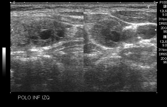
The current scan suggests the thyroid is positioned top-center. The patient is 49, female from Colombia. Observations: predominantly solid composition, isoechogenicity, well defined margins, microcalcifications, tirads 4b. This is an ultrasound image labeled "POLO INF IZQ," suggesting a study of the left lower pole, likely referring to the axilla or perhaps another superficial area.

1. Soft tissue ultrasound using a high-frequency linear transducer in B-mode.

2. Visible Organs and Structures: The image likely displays soft tissue structures beneath the skin. No specific organs are clearly identifiable given the labeling and the typical view of superficial soft tissue images.

3. Features of Each Structure:
 - Size: Specific measurements are not noted, but the image appears to focus on structures typical to a cross-section in soft tissue.
 - Shape and Texture: Several circular to oval structures are visible, which may represent lymph nodes or other soft tissue elements. These structures display a typical hypoechoic center with a possible echogenic hilum.
 - Borders: Generally well-defined, indicating regular soft tissue practice.
 - Symmetry: Not applicable given the image orientation.

4. Additional Findings:
 - Fluid Presence: No clear evidence of free fluid collection in these soft tissue images.
 - Artifacts: There is some posterior enhancement typical under hypoechoic structures.
 - Masses/Lesions: The structures observed do not indicate masses with irregular borders; they are likely normal anatomical features.
 - Vascular Features: Not directly assessable without Doppler imaging.

5. Gray Scale and Doppler Features:
 - Hyperechoic/Hypoechoic Regions: Hypoechoic areas are typical for lymph nodes or vessels.
 - Doppler Findings: Not available in this image set-up.

6. Dynamic Features: Not evaluable from a static image.

7. Image Quality and Limitations: Image quality appears good for soft tissue, though limited by the static form and absence of Doppler for vascular evaluation.

8. Patient Demographic Context: Not provided in the image.

9. Impression/Conclusion: The structures appear consistent with normal superficial anatomical findings, possibly lymph nodes, given the location. No abnormal masses or fluid collections are identifiable. Further evaluation with Doppler or clinical correlation might be required to assess vascular structures or lymph node characteristics fully.
|
29
|
ddti__00000028
|
|
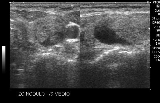
The current scan suggests the thyroid is positioned . The patient is 49, female from Colombia. Observations: tirads 4b, microcalcifications, predominantly solid composition, isoechogenicity, well defined margins. This is a breast ultrasound using a linear transducer in B-mode.

Visible Organs and Structures: The image shows breast tissue with a particular focus on a nodule.

Features of Each Organ/Structure:
- Nodule:
 - Size: The dimensions are not specified in the image but it occupies a significant portion of the visible area.
 - Shape: Appears to be oval and well-defined.
 - Texture (echotexture): The nodule appears hypoechoic compared to the surrounding breast tissue.
 - Borders: The borders are smooth and well-circumscribed.
 - Symmetry: Not applicable as it is a localized finding.

Additional Findings:
- Fluid Presence: No obvious fluid collections are noted.
- Artifacts: No significant artifacts identified.
- Masses/Lesions: The nodule itself is the primary lesion.

Gray Scale and Doppler Features: There are hypoechoic regions indicating the nodule, which may suggest differentials such as a benign cyst or fibroadenoma. No Doppler findings are available for vascular flow assessment.

Dynamic Features: Not applicable in this static image.

Image Quality and Limitations: The image quality is good with clear visualization of the nodule. There is no mention of extraneous factors like bowel gas affecting the image.

Patient Demographic Context: Not specified. Typically, age and sex are crucial for contextual interpretation in breast imaging.

Impression/Conclusion: The ultrasound reveals a well-defined, hypoechoic nodule in the breast, likely representing a benign process such as a fibroadenoma. However, further evaluation with clinical correlation and possibly additional imaging or biopsy may be warranted to rule out malignancy.
|
30
|
ddti__00000029
|
|
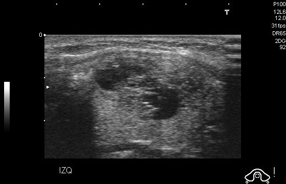
The current scan suggests the thyroid is positioned center. The patient is 29, female from Colombia. Observations: isoechogenicity, well defined margins, microcalcifications, tirads 4a, spongiform composition. The image appears to be an abdominal ultrasound using a linear transducer in B-mode.

Visible organs and structures include the left kidney, which is well-visualized.

For the left kidney:
- Size: The kidney appears normal in size, although no specific measurements are provided.
- Shape: Regular shape observed without deformities or asymmetry.
- Echotexture: Predominantly homogeneous, with areas of hypoechoic regions that may represent the renal cortex.
- Borders: The kidney exhibits smooth and well-defined borders.
- Symmetry: Since only the left kidney is visible, symmetry assessment with the right side is not possible.

There are no significant fluid collections seen. No artifacts such as shadowing or reverberations noted.

Masses/Lesions: No masses or lesions are apparent.

Vascular Features: Doppler evaluation is not visible in this image, so flow patterns and vascular assessments are not applicable.

Dynamic Features: Due to the static nature of this image, dynamic features such as organ motion are not assessable.

Image Quality and Limitations: The image quality appears to be good. There is no apparent impact from factors such as bowel gas or patient positioning.

Patient Demographic Context: Not provided in the image, which limits specific demographic considerations.

Impression/Conclusion: The left kidney shows a normal appearance with no evident abnormalities. Without additional data or the ability to compare bilaterally, this is a limited assessment. Further context or imaging may be necessary for comprehensive evaluation.
|
31
|
ddti__00000030
|
|
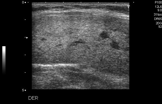
The patient is 44, female from Colombia. Observations: tirads 2, well defined smooth margins. Observed thyroid position: center. The image appears to be an abdominal ultrasound using a linear transducer, likely in B-mode.

Visible Organs and Structures: The image may show soft tissue structures and some subcutaneous tissue layers, but specific organs are not clearly identifiable without more context.

Features of Each Organ/Structure:
- Size: Unable to determine specific sizes of individual structures.
- Shape: Soft tissue appears generally regular, but exact shape is indiscernible.
- Texture (echotexture): The visible tissue shows a heterogeneous echotexture.
- Borders: The boundaries of the visible tissue are not well defined.
- Symmetry: Unable to assess symmetry as bilateral structures are not clearly visualized.

Additional Findings:
- Fluid Presence: No obvious fluid collections identified.
- Artifacts: Some areas of shadowing might be present due to different tissue densities.
- Masses/Lesions: No distinct masses or lesions are visible in this image.
- Vascular Features: Doppler is not used, and no vascular structures are visible.

Gray Scale and Doppler Features: Without specific regions being identifiable as hyperechoic, hypoechoic, or anechoic, clinical significance is limited.

Dynamic Features: No dynamic features apparent, such as motion or changes with respiration.

Image Quality and Limitations: Image quality appears fair, with limitations due to potential shadowing and lack of clear organ structures. Bowel gas or positioning could affect visualization.

Patient Demographic Context: No patient demographic information is available.

Impression/Conclusion: The image captures soft tissue layers without clearly defining specific organs or pathologies, limiting diagnostic interpretation. If an abdominal context is assumed, further imaging or contrasting views are required for better assessment.
|
32
|
ddti__00000031
|
|
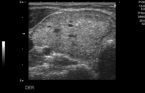
The patient is 44, female from Colombia. Observations: well defined smooth margins, tirads 2. The thyroid gland is located center. Abdominal ultrasound using a curvilinear transducer, B-mode.

Visible Organs and Structures: The image shows the liver, and part of the gallbladder region. The liver appears to be well-visualized, whereas the gallbladder is partially obscured due to positioning or shadowing.

Features of Each Organ/Structure:
- Liver:
 - Size: Difficult to determine precise size from this image.
 - Shape: Appears regular within the section visualized.
 - Echotexture: Generally homogeneous with some scattered hyperechoic areas.
 - Borders: Mostly smooth, but some borders are less defined due to surrounding structures.

Additional Findings:
- Fluid Presence: No significant fluid collections observed.
- Artifacts: Minimal shadowing is noted, possibly obscuring some structures.
- Masses/Lesions: No distinct masses or lesions are identified in this section.
- Vascular Features: Without Doppler, vascular details are not visible in this image.

Gray Scale and Doppler Features: The hyperechoic regions within the liver could indicate the presence of fibrous tissue or other benign processes, but no Doppler findings are available for vascular assessment.

Dynamic Features: Motion cannot be assessed from this single static image.

Image Quality and Limitations: Image quality is fair; evaluation might be limited by shadowing and the partial visualization of the gallbladder.

Patient Demographic Context: Age and sex not provided.

Impression/Conclusion: The liver appears mostly normal within this limited view. No significant abnormalities are noted. The partial visualization of the gallbladder area suggests a need for different positioning or additional images for complete assessment. Further evaluation may involve a Doppler study to assess vascularity and confirm the absence of significant pathologies.
|