| # | key | img1.png | img2.png | txt | img3.png | img4.png | img5.png | img6.png |
|---|
1
|
medpix__00000000
|
|
|
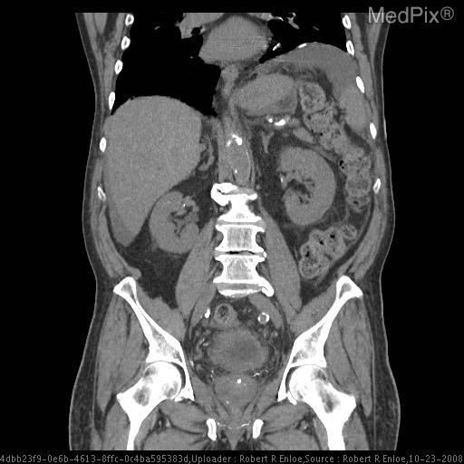
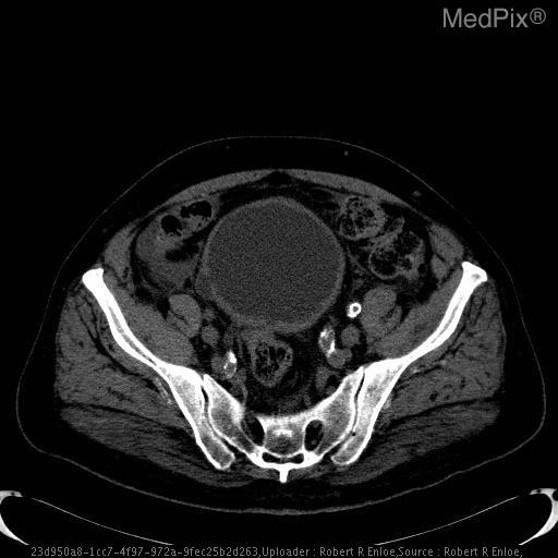
Title: Bladder Diverticulum
History: 73-year-old male with hematuria and numerous white blood cells found on UA
Findings: Bladder with thickened wall and diverticulum on the right.  Diverticulum is mostly likely secondary to chronic outflow obstruction.

Prostate enlargement.
Diagnosis: Bladder Diverticulum
Discussion: Bladder diverticula most often occur as a result of outlet obstruction.  Occasionally, a congenital weakness in the bladder wall adjacent to the ureteral orifice results in a diverticulum.  This is termed a "Hutch" diverticulum.
In children, outlet obstruction causing a diverticulum is rare and can be seen with urethral valves.  In men, diverticula are associated with outlet obstruction from urethral stricture, prostatic hypertrophy, prostatic carcinoma etc.  acquired diverticula are rare in women.
Diverticula usually occur on the lateral bladder walls, rarely the dome.  They are often multiple.  Large diverticula often displace the bladder and or ureters.
diverticula can have wide or narrow necks.  The wide necked variety empty urine readily.  The narrow neck type are slow to empty and therefore are more likely to have urinary stasis.
Infection, tumor and stone formation can occur as a result of urine stasis within a diverticulum.  Tumor formation in a diverticulum is more likely to spread beyond the bladder because the diverticulum wall consists only of urothelium without muscle.
Bladder diverticula can be evaluated with excretory urography, ultrasound, CT and cystoscopy.

Ref:
Dunnick, R., McCallum, R., Sandler, C., Textbook of Uroradiology.
[CT - noncontrast, Coronal] The prostate is enlarged with several calcifications  noted within.  No dominant prostate mass is evident.
[CT - noncontrast, Axial] Bladder is prominent with mildly thickened wall. There is a small posteriolateral diverticulum on the rightward aspect.
|
|
|
|
|
2
|
medpix__00000001
|
|
|
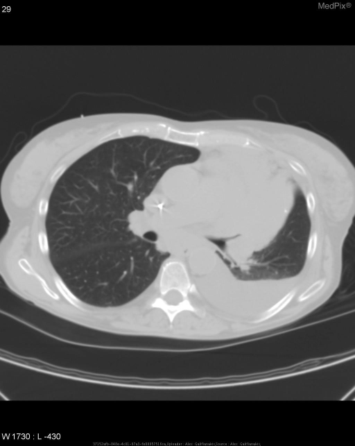
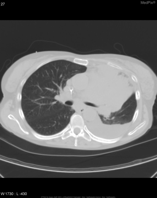
Title: Left upper lobe collapse caused by an enlarging, obstructing small cell lung carcinoma.
History: 60-year-old woman presents with chest pain and shortness of breath.
Findings: •  PA chest radiograph demonstrates left lung volume loss, silhouetting of the left cardiac border, and Luftsichel sign.

•  Lateral chest radiograph shows anterior displacement of the major fissure and elevation of the left mainstem bronchus.

•  CT of the chest reveals an obstructing mass and resultant LUL collapse.
Diagnosis: Left upper lobe collapse caused by an enlarging, obstructing small cell lung carcinoma.
Discussion: Total left upper lobe atelectasis is caused by obstruction of the left upper lobe bronchus.  This may be due to pulmonary mass, mucous plugging or foreign bodies.  In older individuals, a pulmonary mass is a common cause.

PA films of left upper lobe atelectasis demonstrate opacification of the left upper lung field with obliteration of the left cardiac border.  In addition, there is often a relative lucency adjacent to the aortic knob which represents an overinflated lower lobe.  This lucency is referred to as the Luftsichel sign.

The lateral film demonstrates anterior displacement of the major fissure with increased anterior opacification.

This patient had a previously known left upper lobe mass with total left upper lobe atelectasis proven by CT scan.
[CT - noncontrast, Axial] CT of the chest reveals an obstructing mass and resultant LUL collapse.
[CT - noncontrast, Axial] CT of the chest reveals an obstructing mass and resultant LUL collapse.
|
|
|
|
|
3
|
medpix__00000002
|
|
|
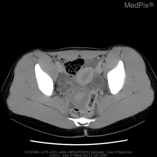
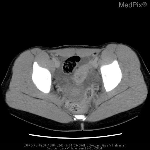
Title: Ovarian torsion
History: 24 hours of pelvic, RLQ pain.
Findings: CT: Large heterogeneous pelvic mass displacing the uterus anteriorly.

US: Enlarged right ovary with central cystic component.  Absent blood flow.
Diagnosis: Ovarian torsion
Discussion: Ovarian torsion is caused by partial or complete rotation of the ovary on its mesenteric pedicle.  This results first in compromise of the lymphatic and venous drainage, causing congestion and edema of the ovarian parenchyma and eventually leading to loss of the arterial perfusion and infarction.  The condition is a surgical emergency much like a testicular torsion.   Torsions may occur in normal or abnormal (ie ovarian cyst, dermoid) ovaries.  This condition usually occurs in children and younger woman who have more mobile adnexa.

The imaging findings of ovarian torsion are variable.  However, any abnormal ovary on ultrasound in the proper clinical setting should suggest the diagnosis.  The sonographic findings depend on the degree of vascular compromise and whether an adnexal mass is present.  The ovary is usually enlarged.  Multiple enlarged cortical follicles in a big ovary with the proper history-acute lower quadrant pain (same side as the abnormal ovary), nausea, vomiting, mildly elevated WBC-is considered a specific sign.  Color and spectral Doppler examination may show absent flow in the affected ovary.  However this is neither a specific nor sensitive sign.  Torsed ovaries may have Doppler flow, and normal ovaries may not show flow on Doppler interrogation.   (Rumack, Diagnostic Ultrasound p.550)
[CT - GI & IV Contrast, Axial] pelvis
[CT - GI & IV Contrast, Axial] pelvis
|
|
|
|
|
4
|
medpix__00000003
|
|
|
Title: Adenocarcinoma of the Lung
History: The patient is a 43-year-old female who presented with a 6-month history of progressively worsening shortness of breath with exertion during her 5-mile runs.  The patient also complained of wheezing but denied having a cough, fevers, chills, or weight loss.  This patient does not have a previous history of any chronic illnesses and does not have any history of tobacco use.
Findings: Chest PA/LAT revealed increased interstitial markings in the right lower lobe.

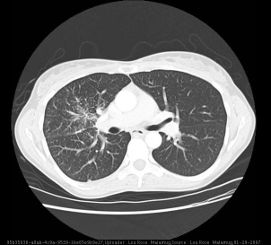
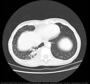
Contrast enhanced chest CT revealed diffuse increased interstitial markings involving the right middle and lower lobes, pleural thickening/scarring of the posterior right lower lobe, and a small right-sided pleural effusion.
Diagnosis: Adenocarcinoma of the Lung
Discussion: Lung cancer has surpassed breast cancer in becoming the leading cause of cancer death in women. Adenocarcinoma accounts for 30-40% of lung cancers and is the most common histologic type.  It is also the most common type of lung cancer in non-smokers (an estimated 10-15% of patients diagnosed with lung cancer are non-smokers).  The most common sites of distant metastases are the brain, bones (most commonly vertebrae), liver, adrenals, and skin.  The American Thoracic Society recommends measurement of a CBC, serum electrolytes, calcium, alkaline phosphatase, albumin, AST, ALT, total bilirubin, and creatinine in all patients with lung cancer.  Abnormal LFTs should be followed up with a contrast-enhanced CT of the liver, and an elevated alkaline phosphatase or calcium level should be followed up with a bone or PET scan to detect metastases (there is conflicting evidence as to which imaging modality has greater sensitivity).  With regard to monitoring response to therapy, one study reported that changes in FDG uptake on PET scan rather than changes in lesion size on CT more accurately correlates with response to therapy.  Stage IV disease is primarily managed with chemotherapy or with palliative measures for symptomatic relief, and the five year survival rate is less than 5%.

Systematic screening for lung cancer is not currently recommended by any major medical organizations.  The USPSTF concluded that there is currently insufficient evidence to argue for or against screening asymptomatic individuals for lung cancer with either chest x-ray, low dose computerized tomography, sputum cytology, or a combination of these tests. Studies evaluating the benefits of screening have shown increased detection of early stage lung cancer in asymptomatic individuals but have not shown a reduction in mortality.  The Prostate, Lung, Colorectal, and Ovarian Cancer Screening Trial is an ongoing trial evaluating the use of a single posterior-anterior CXR, and the Early Lung Cancer Action Project is currently evaluating the use of low-dose spiral CT for screening.
[CT w/contrast (IV), Axial] Contrast enhanced chest CT shows diffuse increased interstitial markings involving the right middle and lower lobes.
[CT w/contrast (IV), Axial] Contrast enhanced chest CT shows pleural thickening/scarring of the posterior right lower lobe.
|
|
|
|
|
5
|
medpix__00000004
|
|
|
Title: Occipital Condyle Fractures
History: 22yo M transported to the emergency department after a motorcycle accident. The head CT showed multiple intraparenchymal bleeds and small subarachnoid bleed. No other fractures of the cervical spine identified. Patient was monitored non-operatively by a hard collar. He developed a right cranial VI nerve palsy two days after the accident due to hemorrhage related to the fracture.
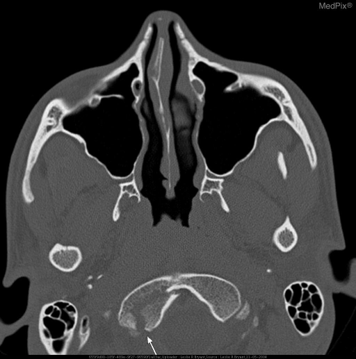
Findings: Axial and coronal CT of the head and cervical spine demonstrating a R Occipital condyle fracture.
Diagnosis: Occipital Condyle Fractures
Discussion: The most common classification is by Anderson and Montesano that describes three basic types of occipital condylar fractures.


Type I is usually the result of an impaction type injury due to asymmetrical axial forces applied to the head and may be seen with other lateral mass fractures in the upper cervical spine.  The result is a comminuted fracture of the occipital condyle at the insertion of the alar and apical ligament complex. It is considered a stable fracture.


Type II is often the result of distraction type forces applied through the apical and alar ligament complex. The result is a basilar skull fracture extending to and involving the occipital condyle with an intact alar and apical ligament complex. It is considered stable unless the fracture completely separates the occipital condyle from the occiput.


Type III is an avulsion type fracture of the occipital condyle. The appearance is a fracture of the distal tip of the occipital condyle resulting in a free condylar fragment that may be displaced into the foramen magnum toward the odontoid process. A type III fracture is considered unstable.
[CT - noncontrast, Axial] Non-contrast CT axial view demonstrates a R occipital condyle fracture at the arrow. There is also a small amount of hemorrhage tracking posterior to the fracture.
|
|
|
|
|
6
|
medpix__00000005
|
|
|
Title: COPD and chronic ILD secondary to leukemia with acute PAP
History: 74 yo female with history of leukemia and COPD now with acute onset of shortness of breath.
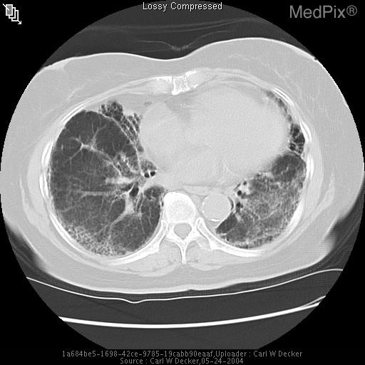
Findings: Predominately basilar and peripheral interlobular septal thickening with scattered areas of ground glass opacity, consolidation, and fibrosis.
Diagnosis: COPD and chronic ILD secondary to leukemia with acute PAP
Discussion: INTERSTITIAL LUNG DISEASE --- The lung interstitium is composed of three components: bronchoarterial (axial), peripheral and parenchymal (using the terminology of Fraser and Pare). The axial component consists of the bronchovascular couplets (bronchiole and accompanying pulmonary artery). The peripheral space is composed of pulmonary veins and paired lymphatics and run within the interlobular septa. The peripheral component is continuous with the pleura. Finally, the interstitial parenchyma consists of the gas exchanging components of the lung (i.e. the walls of the alveoli). An understanding of these components allows one to think logically of the disease categories which affect the lung interstitium. This is especially true is regard to the interpretation of HRCT.

While there are numerous methods by which to categorize ILD none are completely satisfying for ILD consists of a broad array of processes that share similar histopathologic and radiographic manifestations. Broadly they may be categorized as follows: (1) interstitial pneumonias (UIP, DIP), (2) diffuse infiltrative processes characterized by granuloma formation [e.g. sarcoidosis], (3) lymphocytic interstitial pneumonia, (4) pneumoconioses, (5) ILD associated with cysts [LAM and EG], (6) disorders characterized by interlobular septal thickening (pulmonary edema, pulmonary alveolar proteinosis and primary lymphangitic carcinomatosis), (7) eosinophilic syndromes, (8) pulmonary hemorrhage syndromes and (9) vasculitis. The combination of radiographic, clinical and laboratory findings are usually necessary to make a diagnosis and biopsy may be necessary to be definitive.

PULMONARY ALVEOLAR PROTEINOSIS ---- PAP was shown to complicate the underlying COPD in the case presented. As noted above, PAP is within the category of diseases associated with interlobular septal thickening. Populations at risk for development of PAP include the immunocompromised (e.g lymphopenia in children and leukemia in adults); humans exposed to high concentrations of silicon dioxide; those infected with a variety of microorganisms (especially Nocardia, Aspergillus and Cryptococcus).

PATHOLOGIC FINDINGS IN PAP show the alveoli to be filled with fine granular material which stains positive with periodic acid-Schiff stain (PAS+). Further histopathologic findings which may be present include: air-spaces filled with cholesterol crystals as well as needle-shaped crystals and laminated bodies (probably cellular fragments) and degenerating macrophages. Rarely interstitial fibrosis is present.

RADIOGRAPHICALLY PAP was first described in a group of 27 patients in 1958. All demonstrated perihilar vaguely nodular air-space consolidation on plain film.  Less severe disease may manifest as ground-glass opacity only. In general, findings may range from ill defined nodular densities or patchy areas of confluence confined to the lower lung to diffuse consolidation throughout both lungs. On conventional CT PAP may manifest as ill defined consolidation, while on HRCT the characteristic “crazy-paving” may be evident. This latter finding is secondary to a fine linear pattern forming (3-10mm) polygonal shapes superimposed on a background of GGO. Of special note is the fact that “crazy-paving” is NOT pathognomonic of PAP. Differential considerations include BAC, lipid pneumonia, pulmonary hemorrhage, hydrostatic and permeability pulmonary edema, and bacterial pneumonia.
[CT - noncontrast, Axial] CT (3mm collimation) shows peripheral interlobular septal thickening with scattered focal consolidation and GGO.
|
|
|
|
|
7
|
medpix__00000006
|
|
|
Title: Congenital Cholesteatoma
History: 4 year old male with presented with white mass behind the left tympanic membrane noted on routine physical exam.  Pt has no hx of ear surgery, perforations or trauma.
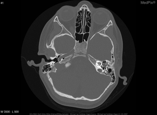
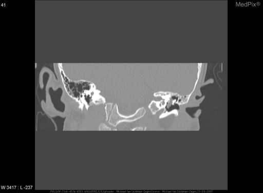
Findings: - Cholesteatoma of the left mesotympanum and epitympanum
- Extends medially toward the malleus and incus without clear erosion of the bones
- No dehiscence of the facial nerve noted
Diagnosis: Congenital Cholesteatoma
Discussion: Congenital cholesteatoma

Cholesteatomas can be divided into two catagories, aquired cholesteatomas caused by traumatic, infectious or post surgical causes and congenital ectopic tissue rests.

Clinical criteria: pearly white mass medial to an intact tympanic membrane, a normal pars tensa and flaccida, and no history of otorrhea, perforation, or previous otologic procedure

Clinical presentation: Most common presentation is a white retrotympanic mass noted on routine otoscopy.  Now typically noted around the age of 4-5 years old.  If they go undetected other symptoms are determined by the middle ear structure they interfere with.  Facial nerve paralysis, conductive or sensorineural hearing loss, vertigo and extension into the middle fossa are all possibilities.  The most common complication is a conductive hearing loss due to fixation of the ossicles by the mass.  However, some large cholesteatomas can allow for sound conduction through the cholesteatoma itself.

Location: Most commonly found in the anterior superior quadrant of the tympanic membrane when found early, but as they grow they tend to migrate to the posterior hypotympanum.

Pathophysiology: There are four theories on how a congenital cholesteatoma is formed, the most widely accepted is the epithelial rest theory
- Epithelial rests: Teed-Michael theory, ectodermal implants have been found in the 10 week old fetus between the first branchial cleft and pouch which is distinct from the surrounding tissue.  They have an unknown function and typically are resorbed at 33 weeks gestion.  If they do not involute then they may form a chronic cholesteatoma.  While there is histologic evidence of rests prior to 33 weeks there are now two documented histological cases such rests persisting after 33 weeks.
- Invagination: This theory suggested that tissue from the external canal invaginates through the tympanic membrane due to a very small inflammatory injury near the neck of the malleus which causes adherence of squamous tissue to the malleus.  This may occur in utero or during childhood.   Another related theory is that there is no actual macroscopic perforation of the tympanic membrane, only microperforations of the basal layer.  This allows for squamous epithelium to enter the middle ear space.
- Implantation: This is actually a theory against the existence of congenital cholesteatomas and implies that they are actually formed by unrecognized perforations of the tympanic membrane.
- Metaplasia: This theory explains that epithelial tissue in the middle ear is via metaplasia to keratinizing squamous epithelium, although this theory appears to be unlikely due to the fact that most congenital cholesteatomas occur in one quadrant of the middle ear.
Histology: Stratified squamous epithelial lined cyst which is filled with keratinous debris, this histology of a congential cholesteatoma is exactly the same as an acquired cholesteatoma.
Radiology: High resolution CT of the temporal bone is the ideal modality for imaging congenital cholesteatomas, especially since most patients are young and without other ear pathology found in patients with aquired cholesteatomas.  On CT a cholesteatoma appears as a hypodense lesion with well defined margins which does not enhance with constrast.
MR imaging of the temporal bone may also be used.  This will show low signal on T1W with moderately high signal on T2W images.  It does not enhance with gadolinium.  MR imaging is useful for differentiating a cholesteatoma from other intracranial processes.

Treatmen
|
|
|
|
|
8
|
medpix__00000007
|
|
|
Title: Pericardial cyst
History: 20 year old healthy male for re-commissioning physical exam.  Patient also C/O worsening cough and congestion over last week
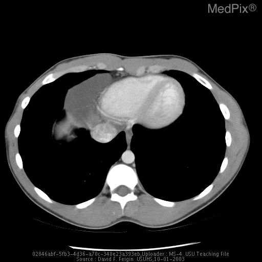
Findings: PA/LAT:  smoothly marginated soft tissue opacity noted in Right cardiophrenic angle, otherwise normal.

CT:  homogenous fluid attenuating and smoothly marginated lesion abutting the right cardiac border with Hounsfield attenuation unit = 7;  Measures 5 x 4.4 x 7 cm
Diagnosis: Pericardial cyst
Discussion: Clinical importance lies in the need to differentiate pericardial cysts from other masses with a similar appearance.  Pericardial cysts represent fluid-filled outpouchings of the parietal pericardium.  They occur in 1/100,000 people.  The cysts rarely calcify and do not communicate with the pericardial space.  One-third of patients are symptomatic at presentation and complain of chest pain and dyspnea.  Pericardial cysts are best diagnosed by CT, MRI, or echo as smoothly marginated fluid-filled structures adjacent to the Rt heart border, although 20% of pericardial cysts lie along the left heart border, sometimes mimicking a prominent left atrial appendage or left ventricular aneurysm.
[CT w/contrast (IV), Axial] CT:  homogenous fluid attenuating and smoothly marginated lesion abutting the right cardiac border with Hounsfield attenuation unit = 7;  Measures 5 x 4.4 x 7 cm
|
|
|
|
|
9
|
medpix__00000008
|
|
|
Title: Jefferson Fracture
History: 27 year old female involved in high speed motor vehicle accident.
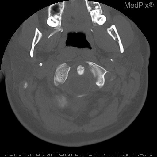
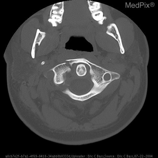
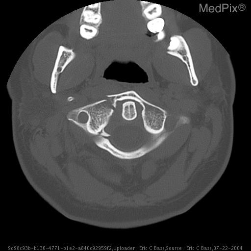
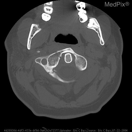
Findings: Selected axial CT images in bone window show multiple fractures of C1 including bilateral anteriolateral fractures with no displacement of left lateral mass and moderate displacement of the right lateral mass best visualized on the coronal reformatted images.  There is also a comminuted fracture in the right posteriolateral portion of the c1 ring with a small, triangular fragment abutting, but not compromising the thecal sac.
Diagnosis: Jefferson Fracture
Discussion: You may search the NIH/NCI cancer treatment protocol database:
http://clinicalstudies.info.nih.gov/

Adult Brain tumors - http://www.cancer.gov/cancertopics/pdq/treatment/adultbrain/healthprofessional

Pediatric Brain tumors - http://www.cancer.gov/cancertopics/pdq/treatment/childbrain/HealthProfessional

Clinical Trials - http://bethesdatrials.cancer.gov/clinical-research/search-results.asp?Specific=31&TextSearch=&Search+for+Clinical+Trials=Search+for+Clinical+Trials
--
[CT - noncontrast, Axial] Selected axial CT images in bone window.
[CT - noncontrast, Axial] Selected axial CT images in bone window.
[CT - noncontrast, Axial] Selected axial CT images in bone window.
[CT - noncontrast, Axial] Selected axial CT images in bone window.
|
|
|
|
|
10
|
medpix__00000009
|
|
|
Title: Horseshoe kidney
History: 78 year old man with new onset of painless hematuria.  There is no significant past medical history.
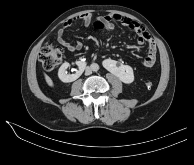
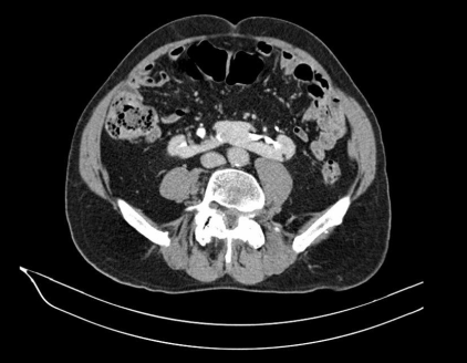
Findings: •  Parenchymal bridge ("isthmus") connecting the inferior poles of both kidneys = Horseshoe kidneys
•  Complete duplication of the right ureter.
•  a 0.7 x 0.3 cm fat containing lesion in the inferior pole of the left kidney.
•  Multiple tiny calcifications bilaterally.
•  1.3cm cyst in the anterior aspect of the left kidney, containing a small calcification on its edge.
Diagnosis: Horseshoe kidney
Discussion: Horseshoe kidney is in a group of congenital kidney anomalies called fusion anomalies.  The fusion occurs at the lower poles in 90% of the cases, while 10% are fused at the upper pole.  This anomaly is found in approximately 1/500-100 people.  It is thought that the abnormal fusion likely occurs when the kidneys are still in the pelvis and the renal capsule is not fully developed.  During the kidneys ascent these fused kidneys are trapped by the inferior mesenteric artery as the isthmus is unable to pass.  The kidneys ascend to their normal positions between the fourth and ninth weeks of gestation.  Complications include: ureteropelvic junction obstruction, recurrent UTI (urine stasis and vesicoureteric reflux), recurrent stone formation, and increased risk of trauma (due to kidney positioning). Horseshoe kidney may occur as an isolated anomaly or associated with other anomalies.  Associated anomalies may include: ureteral duplication, hypospadias, undescended testis, ectopic ureter, anorectal malformations, GI malrotation, Meckel’s diverticulum, neural tube defects, clubfoot, congenital hip dislocation, and ventricular septal defect.  Chromosomal abnormalities have also been associated with horseshoe kidney.  Horseshoe kidney is often evaluated with intravenous urography, CT, and/or ultrasound. MRI and scintigraphy may also be useful.
[CT w/contrast (IV), Axial] There are a few small renal cysts in this Horseshoe kidney.
[CT w/contrast (IV), Axial] There is a small angiomyolipoma in the left portion of the horseshoe kidney - the attenuation is lower than the water in the cysts.
|
|
|
|
|
11
|
medpix__00000010
|
|
|
Title: Hepatic fatty infiltration with focal sparing
History: This is 51 year old woman  with known history of nephrolithiasis and left renal cyst.  She was being followed for stones and had long history of abdominal/pelvic CT’s and KUB films.
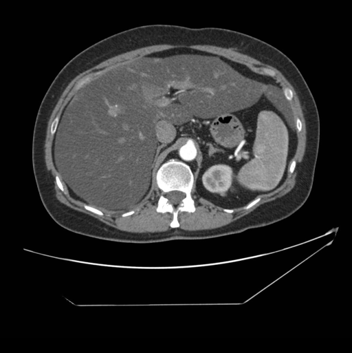
Findings: Selected Images – CT (contrast, arterial phase) of abdomen/pelvis
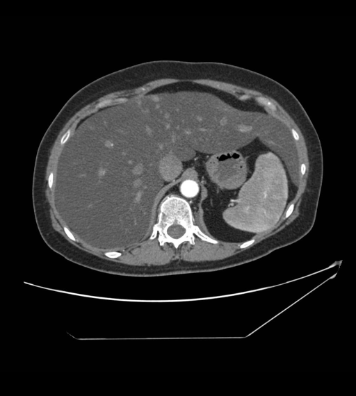
1.  Large, diffusely infiltrated fatty liver with accessory left lobe.  Compare to density of spleen.  Multiple areas of focal sparing in left lobe that appears nodular.
2.  Area of focal sparing near portal vein.
3.  Area focal sparing in left lobe.  Renal cyst and renal calculi.
Diagnosis: Hepatic fatty infiltration with focal sparing
Discussion: Fatty metamorphosis is a common metabolic response of the liver to a variety of inciting agents and disease states.  These agents and states include corticosteroids, chemotherapy, diabetes mellitus, hyperlipidemia, hepatotoxic drugs, obesity, severe hepatitis, chronic alcoholism, malnutrition / malabsorption, jejuno-ileal and jejunocolic bypass surgery, hyperalimentation, congestive heart failure, and blunt hepatic trauma.  Despite these associations, the exact pathophysiologic mechanisms remain obscure.  Possibly diminished portal venous perfusion has a significant effect on producing fatty change and altering hepatic density.  A relative hepatocellular ischemia is proposed to result in decreased parenchymal glycogen and increased accumulations of fat.  The fat content of the liver may rise as high as 40% - 50% of the weight, well above the normal value of approximately 5%.
Regional variation in amount of liver-cell fat can produce entities called pseudolesions or pseudotumors, usually discovered during imaging. Focal fatty sparing in an otherwise fatty liver  occurs in several typical locations: along the gallbladder fossa, along the falciform ligament, occasionally in the periphery of the liver and near the hilum which is perfused with low-insulin blood from a pyloric vein. Because focal fatty sparing can only occur in the presence of fatty liver disease, most of these patients have a history of one of the disease states or agent exposures mentioned above.

Classic features that will help differentiate focally fatty sparing from a hypoechoic mass are that it occurs in these typical locations, often has a geographic (triangular or quadrangular) shape vice being round like most masses and focally fatty sparing, since it is not a mass, will not demonstrate mass effect on adjacent structures, particularly crossing vessels.
[CT w/contrast (IV), Axial] Large, diffusely infiltrated fatty liver with accessory left lobe.  Compare to density of spleen.  Multiple areas of focal sparing in left lobe that appears nodular.
[CT w/contrast (IV), Axial] Large, diffusely infiltrated fatty liver with accessory left lobe.  Compare to density of spleen.  Multiple areas of focal sparing in left lobe that appears nodular.
|
|
|
|
|
12
|
medpix__00000011
|
|
|
Title: Idiopathic Pulmonary Hemosiderosis
History: Patient with diagnosis of idiopathic pulmonary hemosiderosis per open lung biopsy 10/07 who has been relatively asymptomatic but with persistent presence of hemosiderin-laden macrophages in bronchoalveolar lavage and desaturation noted on exercise test in May 2008.

HISTORY
Patient was product of pregnancy complicated by pre-eclampsia. Patient was born at 27weeks after 2 days of antenatal steroids. Significant respiratory distress requiring intubation was noted immediately after birth.

Neonatal course included multiple doses of surfactant. 68 days mechanical ventilation, and 3 months of NICU care. Complications included multiple episodes of pneumonia, presumed sepsis, NEC, anemia, and electrolyte abnormalities.

The patient was discharged home on O2 which she used until 9 months of age, as well as Fe, diuretics, steroids.

After 1 year of age, patient began to have multiple episodes of pneumonia, bronchitis, and asthma requiring hospitalization. Anemia, requiring 2 transfusions, was noted in context of one episode of pneumonia.

In addition, the patient experienced failure to thrive, reaching less than 3% height/weight for her age.

At the present, the patient is at 25% height, 3% weight.

Parents otherwise deny recent URI, cough, wheeze.

Hospitalizations:
after birth x 3 mos
2004-pna
2005-pna and transfusion for anemia
2006-pulmonary hemorrhage, respiratory failure
2007-multifocal pna

Surgeries:
bronchoscopy 12/06, 12/07, 5/08
open lung bx 10/07
port cath placement 12/07

Meds:
Prevacid 15 mg tablet QD
Miralax, 17 gm QD
Iron oral solution75mg/0.6mL- 25 mg/mL
Hydroxychloroquine 25mg/mL- 6ml QD
Tums – 1 tablet QD for calcium
Flinstones MVT
Immunizations: Up to date
Allergies: Cow’s Milk

Developmental History: delayed fine motor skills per MOP; otherwise met all milestones.

Family History: non-contributory

Social History: lives with mother, father, older brother. No pets, no smoking
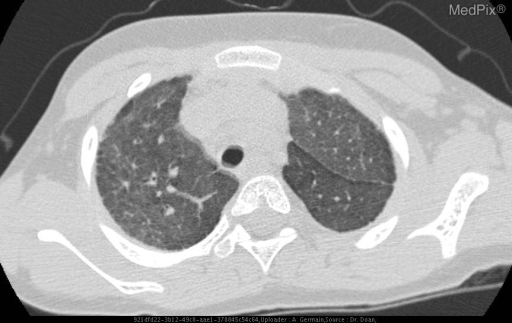
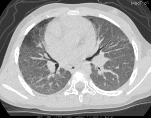
Findings: During acute bleeding episode, areas of increased opacity and decreased lung volume are seen throughout the lungs, especially in the bases.
Diagnosis: Idiopathic Pulmonary Hemosiderosis
Discussion: ***IN PROGRESS****


Lesions/Condition: Idiopathic Pulmonary Hemosiderosis

Cell  of  Origin:

WHO  Grade(s):

Synonyms:

Associations/Predisposing  Factors:

Common  Locations:

Demographics:

Gross  Morphology:

Histology:

Special  Stains:

Gross  Appearance:

Radiology:

Prognosis  and  Treatment:
[CT - noncontrast, Axial] IN PROGRESS
[CT - noncontrast, Axial] IN PROGRESS.
|
|
|
|
|
13
|
medpix__00000012
|
|
|
Title: Pulmonary Embolism
History: Complains of several days of vague abdominal pain, general weakness and shortness of breath.
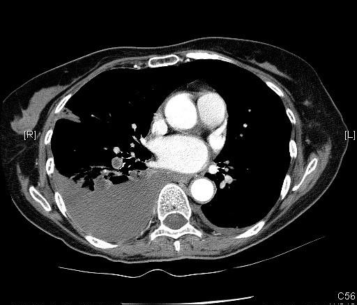
Findings: Bilateral pleural effusions, R>>L.  Right basilar atelectasis.
Main PA filling defect C/W saddle embolism.
Right lower lobe branch of PA filling defect c/w thromboembolism.
Diagnosis: Pulmonary Embolism
Discussion: Pulmonary embolism (PE) can be associated with significant mortality if untreated. The clinical diagnosis of pulmonary embolism is unreliable. Symptoms of PE include tachypnea/dyspnea (most common), tachycardia, hypoxia, pleuritic chest pain, hemoptysis, syncope, and atrial fibrillation. Blood gas may be normal.

The D-dimer blood test is a screening tool for pulmonary embolism. A serum level less than 500 ng/L excludes pulmonary embolism with a 90% accuracy. A positive test is non-specific. Additionally, the test is unreliable in the presence of malignancy, sepsis, recent surgery, or trauma.

The source is  most commonly from deep venous thrombosis in the lower extremity,  however, about 10% arise from clot in the upper extremity.  Risk factors are  Immobilization, Recent Surgery, Underlying Malignancy, History of Deep Venous Thrombosis or Pulmonary Embolism, Estrogen use, or Pre-existing cardiac disease. A low percentage of cases of pulmonary embolism result in pulmonary infarction, due to the presence of the bronchial circulation.

Treatment for PE most commonly consists of anticoagulation with heparin or coumadin. Anticoagulation prevents clot propagation and allows endogenous fibrinolytic activity to dissolve existing thrombiAnticoagulation decreases mortality form 30-60% to <5%.
Thrombolytic agents are not routinely used for the treatment of acute PE. Thrombolytic treatment is generally reserved for patients with massive pulmonary embolism producing circulatory shock (hypotension).

For patients that cannot be anticoagulated, an inferior vena caval filter can be placed in order to prevent life-threatening PE. Major complications occur in about 1% of cases. Complications include central migration of the filter, filter fracture, inferior vena caval perforation, and vena caval thrombosis.
V/Q scanning has been the mainstay for screening symptomatic patients for the presence of pulmonary embolism. A negative V/Q scan essentially excludes PE, and a high probability study is associated with the presence of a PE in about 85% of cases at angiography. Confusion arises with low or intermediate probability examinations, and there is common disagreement among expert readers in the interpretation of scans in these categories. The problem with V/Q scanning is that it does not directly visualize thromboembolism, but rather its effects on perfusion and ventilation . This problem causes the need for probability criteria, which in turn causes confusing results.  Nuclear medicine scanning for PE is probably most useful in previously healthy patients with a normal chest radiograph.   As the complexity of the patients underlying cardiopulmonary disease increases, so does the likelihood that the scan will not be informative (intermediate probability). Using PIOPED criteria, intermediate probability V/Q scans occurred in 60% of patients with COPD, but in only 13% of patients with normal CXR's. However, a generalized abnormality on CXR such as diffuse pulmonary edema or reticulonodular disease may not cause the perfusion lung scan to be abnormal.

The CXR is abnormal in the majority of cases of PE. The PIOPED study showed that among patients with angiographically proven pulmonary embolism, only 12% had chest X-rays interpreted as normal.  (24% of patients with PE in another study had normal CXR's ). Atelectasis and other focal pulmonary parenchymal abnormalities are the most common CXR findings in pulmonary embolism, occurring in up to 68% of patients with PE.  Pleural effusions are also common but usually small and  unilateral. Other palin film findings indicative of PE include regional olig
|
|
|
|
|
14
|
medpix__00000013
|
|
|
Title: Disseminated Coccidioidomycosis
History: The patient is a 23 year old man who presented with four weeks of persistent mid back pain. He received pain medications with minimal relief. His pain gradually worsened over three months to involve his left leg and hip. The patient had associated fevers, weight loss, night sweats, fatigue, and intermittent abdominal pain.
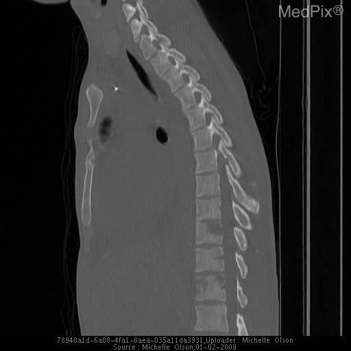
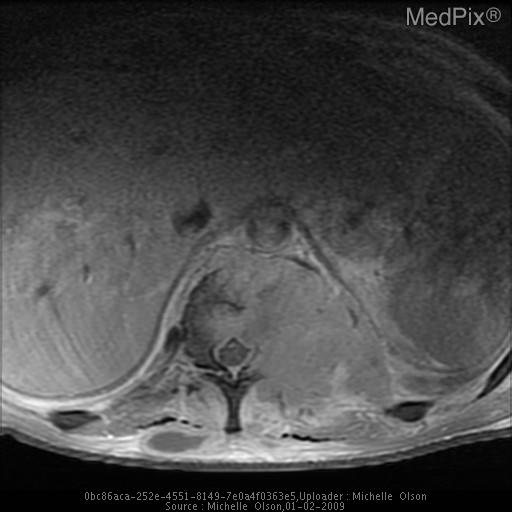
Findings: The frontal and lateral views of the thoracic spine demonstrate a mixed lucent/sclerotic appearance of the T12 vertebral body and the left pedicle.

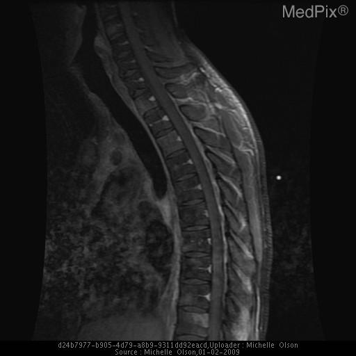
A sagittal view MRI with gadolinium of the thoracic spine and lumbar spine demonstrates enhancing lesions multiple vertebrae, including T12.  Soft tissue enhancement is also present from T9-L2 paraspinal muscles.

A sagittal nonconstrast CT obtained several weeks later demonstrates worsening expansile lytic lesions to T10, T11, and T12.

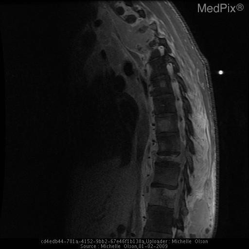
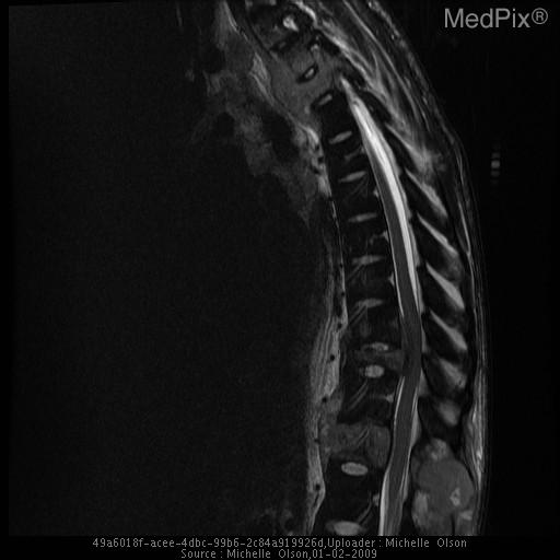
MRI of the thoracic spine was obtained five months after the patient began treatment.  Interval worsening present at multiple levels, including multiple compression deformities and enhancing mass with cortical disruption and contiguous anterior soft tissue mass lifting the anterior longitudinal ligament.  Involvment of the posterior elements is present with narrowing of multiple neural foramina and spinal cord compression.
Diagnosis: Disseminated Coccidioidomycosis
Discussion: Lesions/Condition: Disseminated Coccidioidomycosis

Cell  of  Origin: Dimorphic fungus

Synonyms: Valley Fever

Associations/Predisposing  Factors:  Immunosuppressed or certain ethnic groups

Common  Locations:  Lungs, bone, meninges, skin

Demographics:  Filipinos, Latinos, Asians, African Americans, pregnant women, immunocompromised

Gross  Appearance: Dimorphic fungus

Radiology:  Radiologic findings of coccidioidomycosis disseminated to the skeleton generally involves the axial skeleton. Lesions may be lytic or demonstrate permeative type bone destruction. Lesions on CT may have low attenuation. T1 MRI demonstrates decreased signal and T2 MRI shows increased signal.  Lesions often enhance with gadolinium administration.

Prognosis  and  Treatment:  Patients with disseminated coccidioidomycosis are usually on lifelong antifungal therapy, particularly amphotericin B and the azoles.
[CT - noncontrast, Sagittal] Sagittal CT performed a few weeks later demonstrates worsening expansile lytic lesions to T10, T11, and T12.
[MR - T1W w/Gadolinium, Sagittal] Multiple enhancing lesions in the thoracic vertebrae.  Although T12 is excluded from this image, MR of the lumbar spine demonstrated robust enhancement at T12 as well as multiple lumbar levels.
[MR - T1W w/Gadolinium, Sagittal] MRI obtained five months after the patient began treatment. Sagittal imaging demonstrates more extensive involvement.  T3 vertebral body compression deformity and cortical disruption with a contiguous anterior soft tissue mass lifting the anterior longitudinal ligament.

There is involvement of the T2-3 and T3-4 neural foramina posteriorly. Spinal cord compression is present.

Enhancing foci of multiple other vertebrae. Increased compression of the T10 vertebral body and obliteration of the neural foramina of the T9-10 and T11-12 levels is noted.  An anterior paraspinal mass with anterior cortical disruption and a posterior soft tissue fluid collection at the level of T12.
[MR - T2 weighted, Sagittal] MRI obtained five months after the patient began treatment. Sagittal imaging demonstrates more extensive involvement of T1 and T3. There is a T3 vertebral body compression deformity and cortical disruption with a contiguous anterior soft tissue mass lifting the anterior longitudinal ligament. There is involvement of the T2-3 and T3-4 neural foramina posteriorly. Spinal cord compression is present, best demonstrated on axial images.

Enhancing foci in the anterior T4, posterior T5, and anterior T8 and T9 vertebral bodies. There is increased compression of the T10 vertebral body. There is obliteration of the neural foramina of t
|
|
|
|
|
15
|
medpix__00000014
|
|
|
Title: Chronic Fungal Sinusitis
History: Patient presented after a 9-month cruise - his parents did not recognize his face.  His nose was bigger and his eyes were farther apart than they remembered.  He complained of slight frontal headaches, but had no other symptoms.
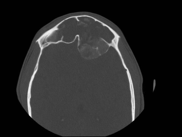
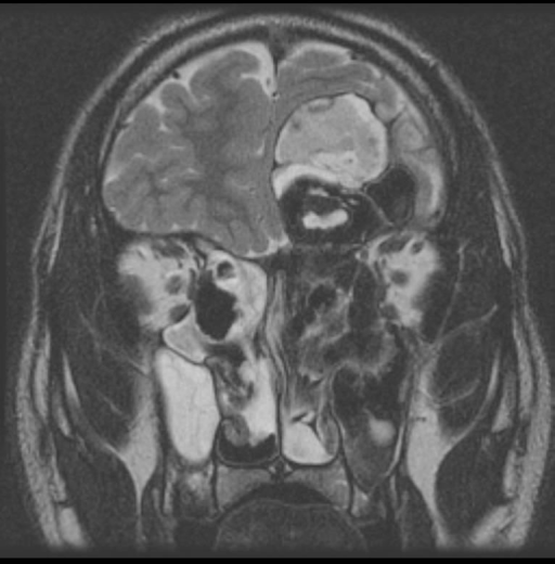
Findings: •  Image 1: Axial CT with expansion and opacification of frontal sinus, with marked thinning and disruption of bone comprising the inner table of the frontal bone
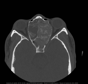
•  Image 2: Axial CT with expansion of ethmoid sinus, with disruption of bone comprising the left lamina papyracea
•  Image 3: Coronal CT with expansion of all paranasal sinuses, with extradural extension into the anterior cranial fossa and extraconal extension into the left orbit
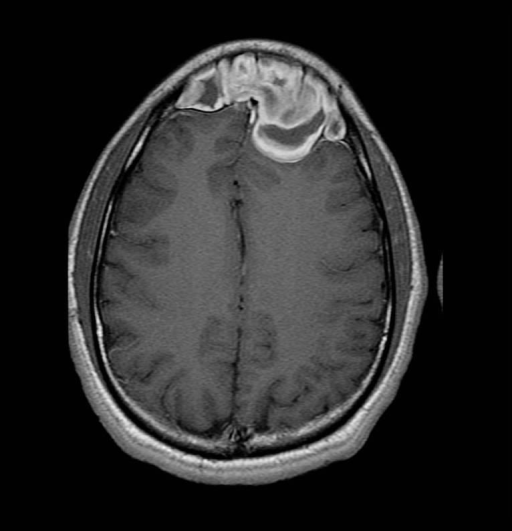
•  Image 4: T1 MRI with heterogeneous signal in the frontal sinuses without changes to the frontal lobe of the brain
•  Image 5: T2 MRI with extradural expansion of heterogeneous mass into the anterior cranial fossa
Diagnosis: Chronic Fungal Sinusitis
Discussion: Lesions/Condition: Nasal sinus aspergillosis

Synonyms: Fungal sinusitis

Associations/Predisposing  Factors: Immunocompromised (invasive fungal sinusitis), Anatomic abnormalities (mycetomas may occur after sinus surgery), Atopic  patients (allergic fungal sinusitis)

Demographics:

   -Mycetoma: Equal in race and gender, may occur at any age
   -Invasive Fungal Sinusitis: Typically immune compromised    adults
   -Allergic fungal sinusitis:  Most common in young adults.  There is a slight male predominance.

Gross Pathology:
  -Mycetoma: Cheesy, semisolid mass
  -Invasive Fungal Sinusitis: Necrotic tissue
  -Allergic Fungal sinusitis: Brownish or green-black thick mucous with "cottage cheese" consistency

Histology:
 -Mycetoma:Fungal hyphae that are tightly packed without allergic mucin.  No tissue invasion.  Calcification can be seen within.
 -Invasive fungal sinusitis: Invasion of mucosa and blood vessels by fungal hyphae. Mucormycosis is the other common organism.
 -Allergic fungal sinusitis: Non-invasive fungi along with eosinophils.  Charcot-Leyden crystals are seen, which are needle-shaped crystalline structures that represent breakdown products of eosinophils.  Other fungi that can also cause: Curvularia, Bipolaris, Fusarium, and Pseudallescheria.

Special  Stains: Gomori methenamine silver


Radiology:

-Invasive Fungal Sinusitis: In patients with invasive fungal sinusitis bone destruction and soft tissue invasion are seen (infiltration of periantral and retroantral fat). On CT bone erosion as well as complete or partial opacification of the affected sinus is seen. CT angiography may reveal vessel involvement (dissection, narrowing, occlusion). Variable signal is seen on T1 and T2.  On post contrast imaging enhancement of the involved tissues is seen.

-Allergic Fungal Sinusitis: On CT, allergic fungal sinusitis typically shows high density material within the sinuses.  On MRI there is low T1 and T2 weighted signal in the sinuses. The peripheral inflamed mucosa will enhance on post contrast imaging.

-Mycetoma: In patients who are otherwise healthy, occasionally a mycetoma may develop. This sometimes occurs afer the chang in the local sinus microenvironment, as can be seen after sinus surgery or radiation therapy. This involves one sinonasal cavity, and on CT is seen as a focal mass within the sinus lumen.  It typically has areas of high density and may have calcification.  The adjacent bone is thick and sclerotic as a result of chronic inflammation. After the administration of contrast, the inflamed mucosa at the periphery of the sinus may enhance.  On MRI, a low signal mass on both T1 and T2 is seen within the sinus.  Occasionally, on T2 the signal may be so low as to be mistaken for air.

Prognosis  and  Treatment:
-Mycetoma: Surgical excision curative
-Invasive Fungal Sinusitis: Radical debridment, IV amphotericin B, with use of itraconazole as long-term suppressive treatment.  Acute fungal sinusitis is life-threatening and can be lethal
|
|
|
|
|
16
|
medpix__00000015
|
|
|
Title: thoracic aortic dissection
History: 76 year-old woman with long history of hypertension, with acute-onset chest pain.
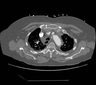
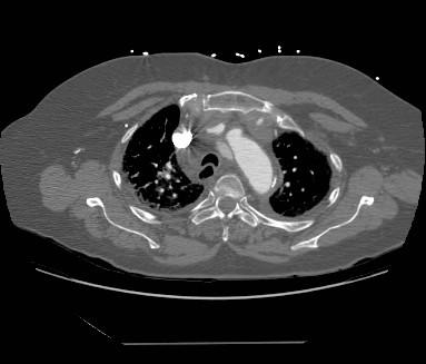
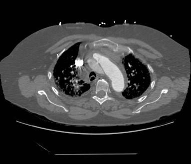
Findings: Multiple axial CT images of the chest with intravenous contrast material demonstrates a Stanford type A thoracic aortic dissection, with a clear intimal flap separating the true and false lumens of the ascending aorta.  Additionally, the pericardium full of dense fluid, likely hemopericardium.
Diagnosis: thoracic aortic dissection
Discussion: Multidetector CT imaging, with faster acquisition of images and utilization of contrast-tracking/event-triggering protocols like CARE Bolus (Siemens) or SmartPrep (GE), is fast becoming the imaging modality of choice for many acute clinical settings such as trauma and the diagnoses of pulmonary embolism as well as aortic dissection.  For the purposes of aortic dissection, the literature advocates at least a four-row CT scanner, with 150 ml dose of non-ionic contrast material intravenously at a rate of 3 ml/sec, with the region of interest for the event-triggering at the level of the thoracic descending aorta.

Aortic dissection is a life-threatening condition that is the most common cause of aortic emergency.  Classification of aortic dissection can be via the DeBakey system, but a simpler Stanford system is now commonly used.  Under the Stanford classification system, a dissection involving the ascending aorta is a type A and any dissection which is limited to the descending thoracic aorta (distal to the left subclavian artery origin) is a type B.  Stanford type A dissections are treated surgically, as these have life-threatening complications such as hemopericardium, dissection which compromises the coronary artery ostia and subsequent massive myocardial infarcts, and severe acute aortic insufficiency causing heart failure and death. Stanford type B dissections, on the other hand, are typically more stable and can be treated medically with close surveillance.
[CT w/contrast (IV), Axial] Multiple axial CT images of the chest with intravenous contrast material demonstrates a Stanford type A thoracic aortic dissection, with a clear intimal flap separating the true and false lumens of the ascending aorta.  Additionally, a pericardial effusion is noted suspicious for hemopericardium.
[CT w/contrast (IV), Axial] Multiple axial CT images of the chest with intravenous contrast material demonstrates a Stanford type A thoracic aortic dissection, with a clear intimal flap separating the true and false lumens of the ascending aorta.  Additionally, the pericardium full of dense fluid, likely hemopericardium.
[CT w/contrast (IV), Axial] Multiple axial CT images of the chest with intravenous contrast material demonstrates a Stanford type A thoracic aortic dissection, with a clear intimal flap separating the true and false lumens of the ascending aorta.  Additionally, the pericardium full of dense fluid, likely hemopericardium.
|
|
|
|
|
17
|
medpix__00000016
|
|
|
Title: Grave's Ophthalmopathy
History: 45 year-old man with a past history of malignant testicular neoplasm and Hashimoto’s thyroiditis, presents now with a several-month history of worsening eye swelling, dryness, and irritation, gradually progressing to lid retraction and intermittent blurry vision, prompting a referral to ophthalmology.  He denies recent trauma, fever, chills, nausea, headaches.
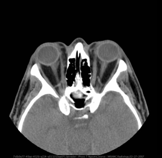
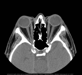
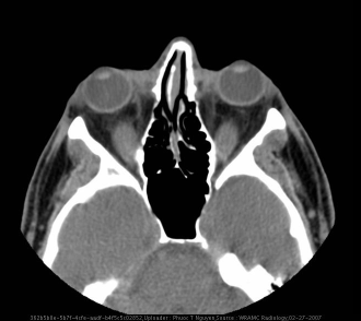
Findings: •  Bilateral proptosis and periorbital tissue edema
•  Enlarged inferior recti muscles
•  Increased periorbital fat distribution
•  Bellies of medial recti enlarged with normal tapering of tendons.
•  Enlarged superior, medial, inferior rectus muscles
Diagnosis: Grave's Ophthalmopathy
Discussion: Grave’s ophthalmopathy (orbitopathy).  Although rare, it can occur in 2% patients with a history of Hashimoto’s thyroiditis.  Grave’s and Hashimoto’s diseases are on opposite sides of the thyroid disease spectrum — manifesting hyperthyroid and hypothyroid symptoms, respectively.  Both diseases can occur in the same person with variable temporal manifestations.  However, the majority of ophthalmopathy cases correlate with Grave’s disease.

Approximately 10% of patients who have ophthalmopathy do not have clinical thyroid disease and are considered euthyroid, but do have elevated serum thyroid autoantibodies and thyroid-specific T-cells circulating.  Within the remaining 90% who do have clinical thyroid disease, approximately 40% have ophthalmic presentation within 6 months after diagnosis of hyperthyroidism, 20% have concurrent clinical thyroid and ophthalmic disease presentation, and 20% have ophthalmic involvement prior to diagnosis of thyroid disease.  Grave’s orbitopathy affects about 16 women and 3 men in a population of 100,000 per year with men having greater severity of disease.

Risk factors for development of Grave's ophthalmopathy are female, positive family history autoimmune diseases, smoking, age, and radioiodine therapy for Grave's hyperthyroidism (compared to surgery or antithyroid drugs).

Grave’s orbitopathy results in bilateral proptosis, lid retraction, chemosis, and vision changes in affected persons attributing to the increase in retroorbital connective tissue and extraocular muscle enlargement.  Complications may involve corneal ulceration and infection due to loss of eye protection and lubrication.

The pathogenesis involves autoantibodies against thyroid stimulating hormone receptor (TSHR) antigens that can be found in the thyroid as well as in other tissues.  It is unknown why retroorbital tissues are predominantly affected.  Studies show that retroorbital fibroblasts and adipocytes express TSHR mRNA suggesting the mechanism of TSHR antibodies directly stimulating the fibroblasts and adipocytes resulting in increased production of glycosaminoglycans (GAGs) and fat tissue.

Another mechanism in the pathogenesis of Grave's ophthalmopathy involves T-cells that recognize self TSHR antigens as foreign.  This autoimmune process stimulates T-cells to produce cytokines such as tumor necrosis factor (TNF) alpha and interferon gamma that recruit lymphocytes and create inflammation and edema within the orbit.  Hence, there is lymphocytic infiltration of extraocular muscles and associated edema accounting for muscle belly enlargement.  It is not known why the inferior recti are usually affected first, followed by the medial recti, then superior recti, and lastly the lateral recti, but this is a consistent finding.

Treatment for Grave's ophthalmopathy depends on severity of symptoms.  If present, treat hyperthyroidism with methimazole.  For mild involvement including dry, irritated eyes, natural tears are helpful during the day and an eye lubricant (methycellulose) is appropriate at night.  For moderate disease that can include conjunctival erythema, progressive proptosis, and diplopia, prednisone 30mg daily for 4 weeks should be initiated additionally.

External radiation may be used in conjunction to predniso
|
|
|
|
|
18
|
medpix__00000017
|
|
|
Title: Acute appendicitis
History: 34 y/o WF w/ abdominal pain for past 30hrs.  Pain started midepigastric and has moved slightly inferior and to the right.  No radiation and no relief with oral meds.  Has had nausea but no vomiting.  Denies fevers, chills, melena, hematachezia, constipation, diarrhea.  Anorexia for 30 hrs. No BM since pain began.  LMP unknown.
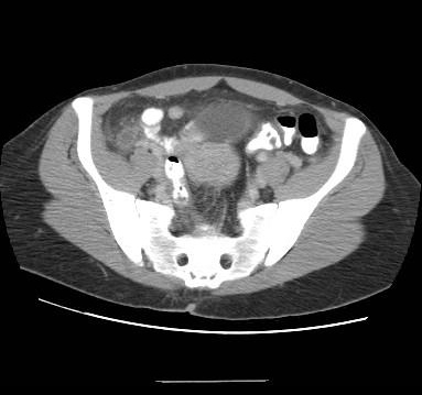
Findings: CT scan abdomen:  Positive “arrowhead sign” in appendix.  Presence of thickened appendix.  Questionable appendicolith.  Terminal ileum normal and seen with contrast,  Good visualization of ileocecal juntion. Cecum normal size without evidence of edema.
Diagnosis: Acute appendicitis
Discussion: In the ED the patient clearly had a diffusely tender abdomen, with evidence of appendicitis.  B-HCG was used to rule out ectopic preg and amylase and lipase were normal.  Likely the patient clinical picture was not overtly obvious for appendicitis (while on paper it seems that way) or else a CT scan should not have been done.  Her CT showed clear evidence of thickening in the appendix with fat stranding.  In addition, there was a positive arrowhead sign, which is indicative of appendicitis.   Finally, the terminal ileum was visible with contrast and showed no evidence of inflammation or changes.  The most likely cause of appendicitis is lymphoproliferation (most common cause of appendicitis) or a fecolith.  The path report is still pending.
[CT - GI & IV Contrast, Axial] CT scan abdomen:  Positive “arrowhead sign” in appendix.  Presence of thickened appendix.  Questionable appendicolith.  Terminal ileum normal and seen with contrast,  Good visualization of ileocecal juntion. Cecum normal size without evidence of edema.
|
|
|
|
|
19
|
medpix__00000018
|
|
|
Title: Typical Bronchial Carcinoid
History: 66yo asymptomatic female with significant past smoking history and abnormal, incidental chest finding on routine EBCT.
Findings: PA/Lat CXR:  There is irregularity to the contour of the right hilum on the PA view.  On the lateral view, there is a central, RML nodule which when going back to the PA, can barely be seen.

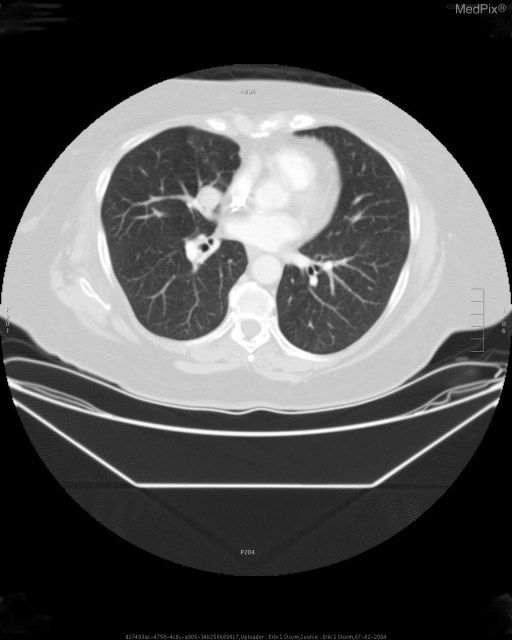
Axial CT:  Note the round, smooth nodule just anterior to the takeoff of the medial RML bronchus.  There is no calcification, and no strong enhancement with intravenous contrast.  The lesion does not appear to involve the bronchial lumen.

Coronal CT reformation:  Round nodule superior to the RML bronchus again demonstrated.
Diagnosis: Typical Bronchial Carcinoid
Discussion: Carcinoids are neuroendocrine neoplasms that are considered malignant based on potential for metastasis (15%)—typically to the liver, bone, adrenals, and brain.  While the vast majority of carcinoids arise in the GI tract (~90%), bronchial carcinoid accounts for 1-2% of all lung neoplasms. These lesions are classified from low grade (typical) to high grade (atypical).  Both extremes have similar imaging features, with the majority of lesions being centrally located, well-defined, and round-to-ovoid in shape.  Calcifications are common; there is no association with tobacco use.  Typical age at presentation is wide, with a mean of 45 years, affecting males and females equally.

Approximately 20% of bronchial carcinoids arise peripherally, distal to the segmental bronchi.  The majority of these neoplasms are of the atypical subtype.  Both typical and atypical subtypes can be associated with hilar and mediastinal lymphadenopathy; hyperplasia results from repeated post-obstructive infections or metastasis.  Local nodal metastasis is more common in atypical carcinoids.

Given the majority of bronchial carcinoids arise within or adjacent to the central bronchi, the most common presenting symptoms are secondary to obstruction with resultant infection or air-trapping.  The lesions tend to be vascular, and hemotypsis is the presenting symptom in ~50%.  Up to 25% of patients are asymptomatic.

Prognosis is dependent on the subtype, with typical carcinoids having 92% 5-year and 76% 15-year survival rates.  Atypical lesions carry a somewhat more dismal 69% and 52% 5/15-year survival rate.  Treatment is by surgical excision.
[CT w/contrast (IV), Axial] 1.6 x 1.8cm right middle lobe nodule, adjacent/anterior to the medial segment bronchus.
|
|
|
|
|
20
|
medpix__00000019
|
|
|
Title: Takayasu"s Arteritis
History: 22 y.o. woman with hypertension.
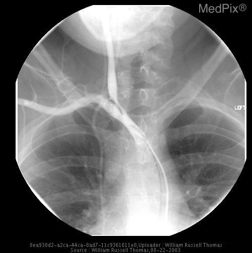
Findings: ABDOMINAL AORTA:  There is narrowing of the abdominal aorta, both above and below the renal arteries.  The superior mesenteric artery is occluded at its origin, and not seen on the lateral view.  There is a large Arc of Riolan from the IMA, which reconstitutes the SMA distribution.  The celiac axis is patent, however, there is a 50% stenosis at the origin.  There are single renal arteries bilaterally, both of which demonstrate stenoses.  On the right, there is a long segment stenosis with approximately 50% narrowing.  On the left, there is 60% to 70% stenosis at the origin of the vessel, extending to an early bifurcation, with an early upper pole branch.  This is also stenotic at its origin.
Diagnosis: Takayasu"s Arteritis
Discussion: DEFINITION
Takayasu’s arteritis refers to a chronic systemic granulomatous vasculitis primarily affecting large arteries (aorta and its branches).

SYNONYMS
Pulseless disease
Aortitis syndrome
Aortic arch arteritis

EPIDEMIOLOGY & DEMOGRAPHICS
•  Most cases have been reported from Japan, China, India, and Mexico.
•  Exact incidence and prevalence is not known.
•  Incidence in the U.S. 2.6/1 million.
•  Females > males 9:1.
•  Seen predominantly in patients <30 yr old.

WORKUP
Any young patient with findings of absence pulses and loud bruits merits a workup for Takayasu’s arteritis. The workup generally includes blood testing to look for signs of inflammation and imaging studies with the angiogram being the diagnostic gold standard.

LABORATORY TESTS
•  CBC may reveal an elevated WBC count
•  ESR is elevated in active disease
IMAGING STUDIES
•  Ultrasound: Carotid, thoracic, and abdominal ultrasound are useful adjunctive imaging studies in diagnosing occlusive disease resulting from Takayasu’s arteritis ( Fig. 1-358 ).
•  Doppler and noninvasive upper and lower extremity studies are helpful in assessing blood flow and absent pulses.
•  CT scan is used to assess the thickness of the aorta.
•  Angiogram can show narrowing of the aorta and/or branches of the aorta, aneurysm formation, and poststenotic dilation.


Angiographic findings are classified as four types:
Type I: Lesions involve only the aortic arch and its branches.
Type II: Lesions only involving the abdominal aorta and its branches.
Type III: Lesions involving the aorta above and below the diaphragm.
Type IV: Lesions involving the pulmonary artery.


PEARLS & CONSIDERATIONS
COMMENTS
Overall the long-term prognosis of treated patients with Takayasu’s disease is good, with >90% of patients surviving more than 15 yr.
[CT w/contrast (IV), Axial] Carotid stenosis
|
|
|
|
|
21
|
medpix__00000020
|
|
|
Title: biliary leak, status post laparoscopic cholecystectomy
History: s/p recent laparoscopic cholecystectomy, now with fevers and abdominal pain
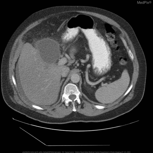
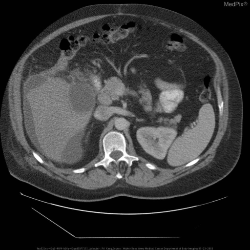
Findings: Axial CT with intravenous and oral contrast material demonstrates a large fluid collection predominantly within the porta hepatis around multiple surgical clips consistent with prior cholecystectomy.  More fluid is seen around the right lobe of the liver and more inferiorly within the right paracolic gutter.  Peritoneal fat around the fluid collection demonstrates stranding suggestive of inflammation.

Planar and SPECT images after the administration of Tc99m-disofenin IV demonstrates large amount of abnormal radiotracer accumulation within the area corresponding to the large fluid collection seen on the CT images, consistent with a biliary leak.  Note that a tubular area of radiotracer uptake leading from the large collection apparently outside the body is the collection within the percutaneous drainage tube placed prior to the scintigraphic study.
Diagnosis: biliary leak, status post laparoscopic cholecystectomy
Discussion: Laparoscopic cholecystectomy (LC) has replaced open cholecystectomy for most routine treatment of gallbladder disease by cholecystectomy due to clear advantages associated with any laparoscopic procedures, namely shortened inpatient stays, smaller incisions yielding better cosmetic results and requiring smaller amounts of pain medication, better pulmonary toilet due to better pain control, and so on. (1, 2)  Early days of LC demonstrated a higher incidence of bile duct injuries and biliary leak, and initially this phenomenon was attributed to the "learning curve", but there is increasing evidence that the "learning curve" may not be entirely the reason why this is so. (2)

In the literature, biliary leaks after LC occur in approximately 1% of patients.  In a series of 64 patients with leaks addressed with endoscopic evaluation (ERCP), the vast majority of the leaks (50) were from the cystic duct.  Other places of biliary leaks were the ducts of Luschka (4), common bile duct (6), and common hepatic duct (4). (5)

Scintigraphic evaluation of patients with potential biliary leaks after LC has been clearly established in the literature (3, 4).  Other anatomic modalities such as US or CT can demonstrate a fluid collection, and a bile leak can be suggested from history, but it would be difficult to actually demonstrate a leak via purely anatomic studies alone.  Scintigraphic evaluation with an agent such as Tc99m-disofenin, as in this case, will clearly demonstrate whether or not the fluid collection is indeed a leakage of bile from the biliary system.

Treatment of biliary leaks in this setting can be done by percutaneous drainage, but the literature demonstrates increasing use of endoscopic evaluation, sphincterotomy, and possible stent placement.  The rationale is that by decreasing the resistance of the normal flow through the common bile duct and into the duodenum, the biliary leak will, in time, close itself.  Percutaneous drainage may be appropriate for patients with possible infection of the bile collection within the peritoneal cavity, which was suspected in this case. (1, 2, 5)
[CT w/contrast (IV), Axial] Axial CT with intravenous and oral contrast material demonstrates a large fluid collection predominantly within the porta hepatis around multiple surgical clips consistent with prior cholecystectomy.  More fluid is seen around the right lobe of the liver and more inferiorly within the right paracolic gutter.  Peritoneal fat around the fluid collection demonstrates stranding suggestive of inflammation.
[CT w/contrast (IV), Axial] Axial CT with intravenous and oral contrast material demonstrates a large fluid collection predominantly within the porta hepatis around multiple surgical clips consistent with prior cholecystectomy.  More fluid is seen around the right lobe of the liver and more inferiorly within the right paracolic gutter.  Peritoneal f
|
|
|
|
|
22
|
medpix__00000021
|
|
|
Title: L parietal ischemic infarct
History: 74 yo WF with Diabetes and HTN, S/P cystectomy, TAH/RSO and ventral hernia now with acute mental status changes 2 days post op.  On first night postop, patient had a one-hour episode of hypotension (SBP 80-90)
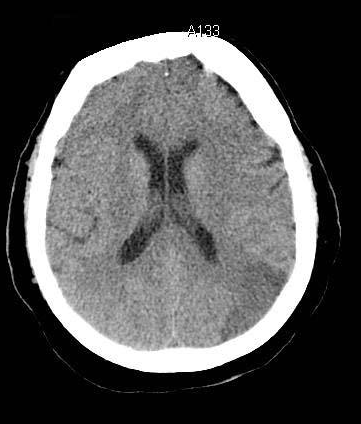
Findings: Noncontrast CT shows hypodensity in L parietal lobe with no visible compression of other structures.  No hyperdens or hemorrhagic areas are noted.

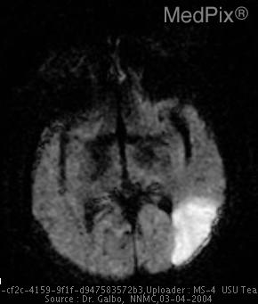
MRI DWI image shows hyperintense signal within same area demonstrating restricted diffusion of water molecules.
Diagnosis: L parietal ischemic infarct
Discussion: CT has been recognized as the standard of care for initial imaging of suspected strokes.   It is able to discriminate between an ischemic vs a hemorrhagic cerebral infarct.  However, it may be relatively less sensitive during the first 12 hours and is not consistently high until after 24 hrs.

MRI may be needed - especially with Diffusion Weighted Imaging has been studied as a predictor of early edema due to cytoxicity to neural cells.  If edema is present, a hyperintense signal will be present in the affected area due to restricted diffusion of water molecules. For the same reason, these areas will have correspondingly low signal on ADC (Apparent Diffusion Coefficient) images.
[CT - noncontrast, Axial] Noncontrast CT shows hypodensity in L parietal lobe with no visible compression of other structures.  No hemorrhage noted.
[MR - DWI Diffusion Weighted, Axial] DWI image from MRI shows hyperintense signal within same area demonstrating restricted diffusion of water molecules.
|
|
|
|
|
23
|
medpix__00000022
|
|
|
Title: Atypical Pulmonary Hamartoma
History: 58 yo man presented with neck pain to his primary care physician.  An MRI of the cervical spine was obtained revealing an abnormal finding.
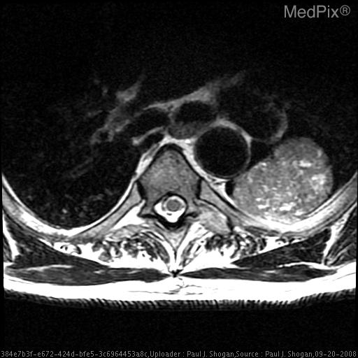
Findings: T2 weighted axial MR image reveals a well-marginated mass with smooth borders in the left lung at the level of the aortic arch, which demonstrates multiple small foci of increased T2 signal intensity, giving the mass a speckled appearance.

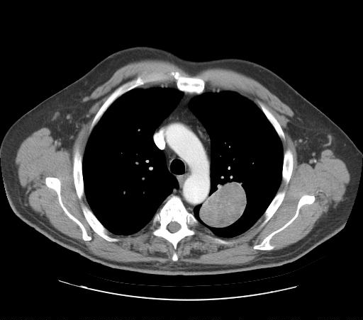
Contrast enhanced axial CT image shows a homogeneous, well-marginated, solid mass with minimal contrast enhancement in the left upper lobe.  No fat or calcification is evident.
Diagnosis: Atypical Pulmonary Hamartoma
Discussion: Lesions/Condition:   Pulmonary Hamartoma

Predisposing Factors:

Most commonly occurs in the fourth to fifth decade.  Most common chromosomal abnormality involves the q13-q15 region of chromosome 12.

Symptoms:

Most patients presenting with peripheral pulmonary hamartomas are asymptomatic.  When symptomatic, hemoptysis is the most common presenting symptom.

Discussion:

Pulmonary hamartomas are benign neoplasm with 90% arising within the lung parenchyma, and are usually in a peripheral location.  They comprise approximately 5% of all solitary pulmonary nodules.  Pulmonary Hamartomas contain cartilage surrounded by fibrous connective tissue with variable amounts of fat, smooth muscle, and seromucous glands.  Approximately 30% contain calcium in the form of multiple clumps (‘popcorn’ calcification).

Generally, pulmonary hamartomas can be observed by CT alone.  Indications for transthoracic biopsy or resection are rapid growth, a size greater than 2.5 cm, or new pulmonary symptoms.

Radiology:

Chest Radiographs:
Well-circumscribed, smoothly marginated solitary nodule without lobar predilection.  Most pulmonary hamartomas are  smaller than 4 cm in diameter.  Calcification is visible in less than 10% of cases.

CT:
Well-circumscribed nodule with a smooth or lobulated border.
Composed entirely of fat, a mixture of fat and soft tissue, or fat and calcification.  Calcification when present is in the form of multiple clumps of calcium dispersed throughout the lesion (“popcorn” calcification)
[CT w/contrast (IV), Axial] Contrast enhanced axial CT images show a homogeneous, well-marginated, solid mass with minimal contrast enhancement in the left upper lobe.  No fat or calcification is evident.
[MR - T2 weighted, Axial] T2 weighted axial MR image reveals a well-marginated mass with smooth borders in the left lung at the level of the aortic arch, which demonstrates multiple small foci of increased T2 signal intensity, giving the mass a speckled appearance.
[MR - Other Pulse Seq., Coronal] 3D plane localizer MR image reveals a mass in the left lung.
|
|
|
|
|
24
|
medpix__00000023
|
|
|
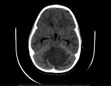
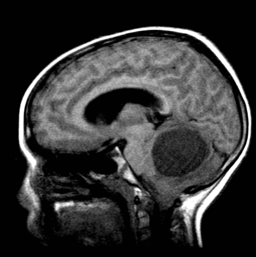
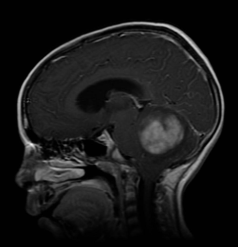
Title: Pilocytic astrocytoma
History: His mother provides a history of increasing headaches over past month, gait disturbances, and possible seizure activity.
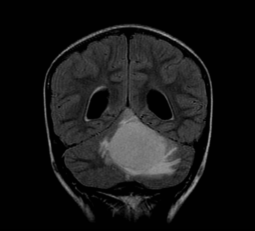
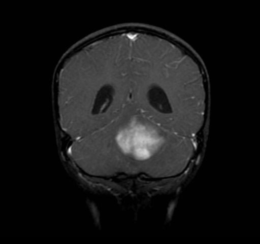
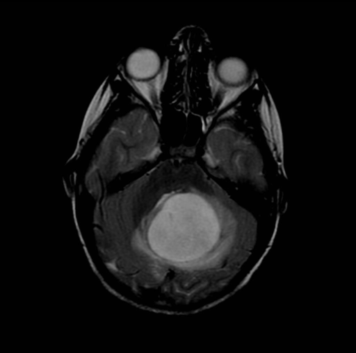
Findings: Large, solid mass in the left cerebellar hemisphere with ill-defined margins and significant surrounding T2 signal abnormality representing vasogenic edema.  This exerts mass effect upon and displaces the 4th ventricle.  Mild hydrocephalus.  Diffuse, heterogeneous enhancement following IV gadolinium infusion.  CT scan demonstrates a notably low attenuation mass, and inferior displacement of the cerebellar tonsils to the level of the foramen magnum.
Diagnosis: Pilocytic astrocytoma
Discussion: Cerebellar  astrocytoma  accounts  for about 11 - 30% of primary CNS tumors in children, and has a post-resection survival of up to  94% at 10 years.  This tumor makes up about one-third of childhood posterior fossa tumors, typically presenting  late  in  the  first decade  or  in  the  early second decade.  These well-circumscribed masses are often completely  resected surgically,  with  no  other therapy required.

Cerebellar  astrocytoma  tends  to involve the hemispheres, although extension into or origin  within  the  vermis  or  even  the fourth  ventricle  have  been  described infrequently.  It may be predominantly cystic with a mural nodule (as shown in a  different patient,  Film .4), multicystic, or predominantly solid (diffuse) on CT or MR.  Recognizable cysts occur in up to 80%.   Histologically, most  are juvenile  pilocytic astrocytomas similar to those of the third ventricle region.   Diffuse  infiltrating  fibrillary  types occur  in  up  to  15%  of cerebellar astrocytomas and  have  a less favorable prognosis.    Frank   malignancy,   leptomeningeal seeding, and spontaneous hemorrhage are distinctly unusual.

Radiographically,  cerebellar  astrocytoma  arises  within  the cerebellar hemisphere and less  often  in the  vermis,  and  displaces the fourth ventricle without invading it.  On CT, 10-25% contain foci of calcification.  The mass is hypointense on T1-weighted  and  hyperintense on  T2-weighted MR, with one or more cystic components.  After contrast administration, the  mural  nodule  and/or  solid  components enhance, rendering nonenhancing cysts more conspicuous.

In  children, vermian or diffuse astrocytoma or those involving the fourth ventricle may mimic medulloblastoma or ependymoma.  In older    adolescents    or    adults,   hemangioblastoma   may   present as a cystic mass indistinguishable from cerebellar astrocytoma.  Metastasis  is  the most  common  posterior  fossa  neoplasm in adulthood.  Rarely vascular malformation,  abscess,  or   lymphoma   might   have   a   similar radiographic appearance.
[CT - noncontrast, Axial] Mid-posterior fossa axial CT slice demonstrates large hypoattenuating mass and vasogenic edema displacing the 4th ventricle anteriorly and to the right.
[MR - T1W - noncontrast, Sagittal] Axial mid-sagittal T1 demonstrates large hypointense mass displacing and exerting mass effect upon the 4th ventricle - causing inferior displacement of the cerebellar tonsils into the foramen magnum.
[MR - T1W w/Gadolinium, Sagittal] Axial mid-sagittal post-gad T1 demonstrates large, heterogeneously-enhancing solid mass with poor definition of borders.
[MR - FLAIR, Coronal] Coronal T2 FLAIR through posterior fossa demonstrates large mass with surounding vasogenic edema.
[MR - T1W w/Gadolinium, Coronal] Coronal post-gad T1 demonstrates heterogenously-enhancing solid posterior fossa mass with somewhat indistinct margins.
[MR - T2 weighted, Axial] Axial T2 images through the posterior fossa demonstrates hyperintense a solid mass, vasogenic edema, and displacement of the 4th ventricle.
|
|
|
|
|
25
|
medpix__00000024
|
|
|
Title: Cerebral infarct, PCA territory, bilateral
History: Sudden onset of blindness in both eyes
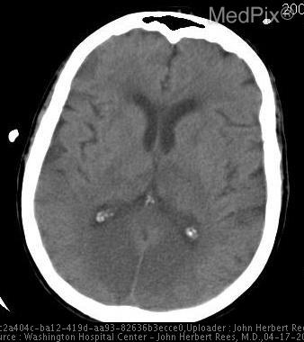
Findings: Bilateral occipital lesions of abnormal decreased attenuation
Diagnosis: Cerebral infarct, PCA territory, bilateral
Discussion: Thromboembolic disease is, by far, the most common cause of acute stroke accounting for 4 out of 5 patients who present with cerebral infarct.  Cerebral hemorrhage is the next most common with almost 1 in 5 patients.  Venous occlusive disease is the next, but much less frequent than the previous mentioned etiologies.

The two most common causes of thromboembolic disease are atherosclerosis and cardiac origin.  Atherosclerosis follows a typical distribution of accumulation within the arterial distribution of the brain.  The internal carotid artery is first, then the distal basilar artery, carotid siphon, and middle cerebral artery.  The parts of the brain perfused by the middle cerebral artery and basilar artery are most vulnerable to infarct due both the region of atherosclerotic disease and flow patterns that cause more proximal sources of thrombus to favor those vessels.

The first patient’s infarct is due to occlusion of penetrating vessels of the right middle cerebral artery.  Likely the lenticulostriates which supply the lentiform nucleus, caudate capsule, and internal capsule, as well as the thalamoperferators were affected.  These types of strokes, when smaller than 15mm, are called lacunar infarcts and they account for 20% of all strokes.  Patients with lacunar infarcts can present with a pure motor hemiparesis like this patient or a pure hemisensory deficit, hemiparetic ataxia, dysarthria, or hand deficit.

CT examination early in the course of an acute stroke can be negative and cannot exclude this diagnosis.  Stroke in the emergent setting is more a clinical diagnosis and if suspected, imaging with MR is prudent.  CT, CTA, MR, and diffusion-perfusion imaging can be useful in management decisions regarding thrombolysis.
[CT - noncontrast, Axial] Bilateral PCA territory infarcts. Notice the abnormal low attenuation in the medial portion of both hemispheres posteriorly (occipital lobes).
|
|
|
|
|
26
|
medpix__00000025
|
|
|
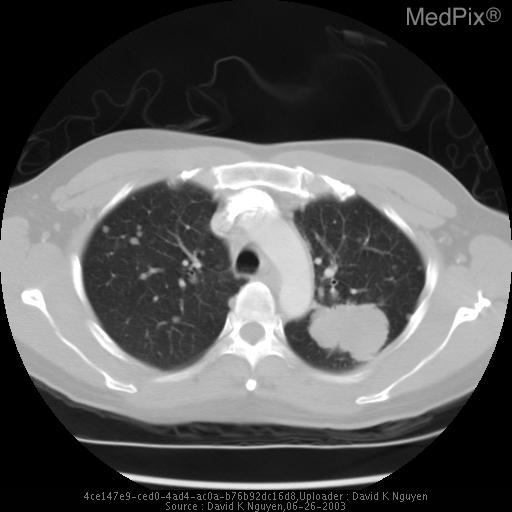
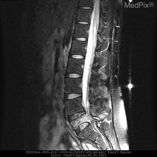
Title: vertebra plana
History: 39 y/o non-smoking female presenting with back pain for two months. No history of fever or weight loss.
Findings: -CT/MRI: There is vertebra plana of L5, with edema and enhancement of the remaining marrow into the posterior elements.  There is displacement of the thecal sac posteriorly by the extruded bony fragments.

-RADIOGRAPH/CT:
Left upper lobe lung mass with numerous bilateral pulmonary nodules, consistent with metastatic disease.
Diagnosis: vertebra plana
Discussion: Marked flattening of a vertebral body is termed vertebra plana.
Langerhans cell histiocytosis is the most common cause of vertebra plana.  Other disease processes should be considered in the differential diagnosis if the clinical situation warrants. Other causes of vertebra plana include:
• multiple myeloma
• metastatic disease
• Ewing sarcoma
• lymphoma
• leukemia
• Gaucher disease
• aneurysmal bone cyst
• trauma
• infection

Because most lesions spontaneously regress, vertebra plana is often treated conservatively. In young patients, vertebral height typically is partially restored with growth of the skeleton. Temporary bracing may help to relieve symptoms.

Radiation may be indicated for treatment of mild neurologic signs. Surgical decompression and fusion with instrumentation is indicated for rapidly progressive neurologic signs or cord compression that is not responsive to radiation.
[CT - noncontrast, Axial] Left upper lobe lung mass with numerous bilateral pulmonary nodules, compatible with metastatic disease.
[MR - T2 weighted, Lateral] There is vertebra plana of L5
|
|
|
|
|
27
|
medpix__00000026
|
|
|
Title: Achalasia
History: 33 year-old woman with shortness of breath and dyspnea on exertion.
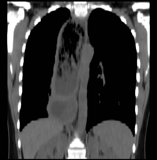
Findings: Frontal chest radiograph demonstrated a wideneded mediastinum on the right side, which on the lateral chest radiograph was a mass within the middle mediastinum superiorly and crossed into the posterior mediastinum inferiorly.

Non-contrast CT examination of the chest demonstrates a diffusely dilated esophagus filled with mottled soft tissue consistent with ingested food material.

Imaged from an upper GI fluoroscopic examination demonstrates a dilated thoracic esophagus with a focal narrowing at the gastroesophageal junction.  A complete lack of peristalsis was noted during the examination.
Diagnosis: Achalasia
Discussion: Clinical symptoms include dysphagia, foul breath, regurgitation and/or aspiration. The condition is usually insidious, developing between age 30-50, and is likely due to a defect in esophageal innervation, with impairment or absence of ganglion cells in Auerbach's Plexus.
    Radiographically, abnormal peristalsis (often completely absent) and abnormal LES opening are seen with barium swallows. Barium remains above the LES until pressure  (hydrostatic or nonperistaltic contractions) wedges the LES open. When this occurs, the barium column appears as an elongated "V"  or "bird beak" yet latter term is not specific for achalasia.
    Upper endoscopy is necessary to rule out a benign or malignant stricture. Failure of LES relaxation is confirmed by esophageal manometry that also reveals complete absence of contractile activity, referred to as aperistalsis (“classic achalasia”) or persistent spastic contractions (“vigorous achalasia”).
    Treatment options aim to relax the LES, and include medicines (smooth muscle relaxants), balloon dilation (70-80% success, often requires re-dilation, risk of perforation), or surgical myotomy of the LES (now often done laparascopically, and combined with antireflux surgery.)
[CT - noncontrast, Coronal] Coronal reconstruction of the axial non-contrast chest CT demonstrates a diffusely dilated esophagus filled with mottled soft tissue consistent with ingested food material.
|
|
|
|
|
28
|
medpix__00000027
|
|
|
Title: Lung Metastasis
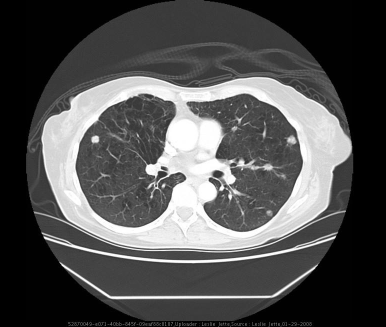
History: 57yo F with 1 mo cough presents for f/u CXR. CXR 1 mo ago showed interstitial infiltrates and was prescribed abx.  PMH includes colon cancer – dx 5y ago as stage 2 with hepatic metastases.
Findings: Numerous, scattered, small masses
Diagnosis: Lung Metastasis
Discussion: Cancers can metastasize to the lungs by several pathways.  Most commonly, cancers spread to the lungs via pulmonary arteries or lymphatic channels.  Less often, spread is by direct extension or endobronchial spread.  Cancers that commonly metastasize to the lungs include:  prostate, breast, renal, thyroid, testicle, gastrointestinal, melanoma, and sarcomas.
Radiograpically metastases tend to be multiple (95%).  They are more often in the lung periphery and bases.  Typically they have sharp margins.  They can cavitate, particularly squamous cell carcinomas.  Some may have calcifications.
[CT w/contrast (IV), Axial] Multiple small, scattered masses
|
|
|
|
|
29
|
medpix__00000028
|
|
|
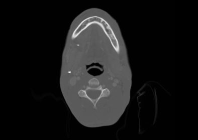
Title: Sialolithiasis resulting in Sialoadenitis
History: Hx not provided
Diagnosis: Sialolithiasis resulting in Sialoadenitis
Discussion: Sialolithiasis is an accumulation of hardened deposits in the salivary ducts. The most common site of occurrence is in the submandibular  (Warthon’) duct  which accounts for about 80% of all salivary duct stones.1  The large majority of cases occur in patients over 30 years of age with rare occurrences in children. The exact etiology of stone formation in the salivary ducts remains uncertain however it is believed to be the result from trauma, inflammation of the ductal epithelium or other processes which slow the movement of saliva down the salivary ducts resulting in stagnation. Patients typically present complaining of neck swelling and colicky pain, though this patient denied pain. Diagnosis is achieved through direct palpation of the stone and imaging which should include CT with contrast or ultrasound. Sialolithiasis often leads to Sialoadenitis, which is inflammation of salivary gland, caused by granulomatous processes or more typically in the case of Sialolithiasis infection caused by bacterial migration up the obstructed duct.  Staphylococcus, Streptococcus viridans and S. pneumoniae are usually implicated in the infection. 2   Treatment is with broad spectrum antibiotics, sialogogues (sour candy to promote salivation), massage, manual removal of stones near the orifice. 1(p1239),2(p935)  Failure of these treatments should prompt consideration of surgical intervention, which is not considered first line treatment due to the high incidence of post-operative stricture formation and resulting predisposition for chronic Sialoadenitis.1(p1240)
[CT w/contrast (IV), Axial] 4-5 mm calcified stone in the expected region of the right submandibular gland duct. There is a small surrounding focal fluid collection. The right submandibular gland is enlarged in comparison to the left side. The left submadibular gland is normal. There is normal symmetric appearance to the parotid and sublingual glands. Vascular structures are normal. There is no pathologic by size adenopathy in the neck. Osseous structures are normal.
|
|
|
|
|
30
|
medpix__00000029
|
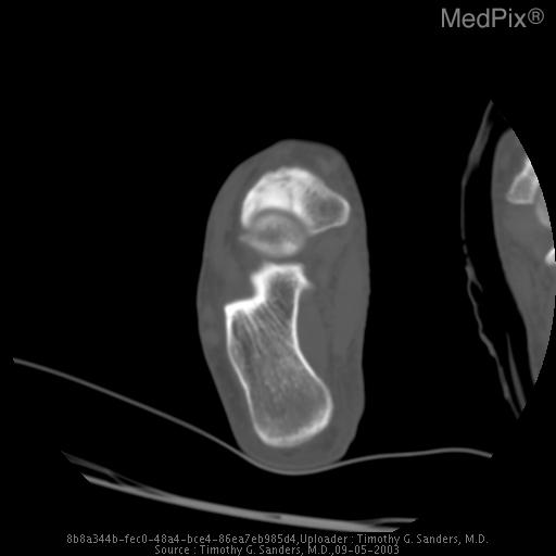
|
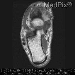
|
Title: Stress fracture Tarsal Navicular Bone
History: 21 y.o. female long distance runner presents with a several week history of pain along the dorsum of the foot. The pain is exacerbated by running.
Diagnosis: Stress fracture Tarsal Navicular Bone
Discussion: Stress fractures occur when normal bone is subjected to abnormal stress or repetitive stress, and they typically occur in athletes who have either recently changed their type of training or have increased their level of training. Bone is a very dynamimc tissue that continuously remodels as a result of altering stresses. Repetitive stress overload results in an imbalance of bone remodeling. The abnormal level of stress results in the bone resorption exceeding the rate of bone repalcement leading to a weakening of the bone.  There is a continuum of injury beginning with weakening of the bone, fatigue, stress fracture and finally, complete fracture of the involved bone.

Stress injuries of bones are very common among athletes and can occur in nearly any bone, however, the vast majority of stress injuries occur in the bones of the lower extremity. Tarsal navicular stress fractures typically occur in elite athletes, including runners, basketball players, gymnasts, and football players. Those athletes that play on artificial turf seem to be at an increased risk for stress fractures of the tarsal navicular bone.

The diagnosis of tarsal navicular stress fracture is often delayed many weeks to months because the onset of symptoms are insidious and in addition, the diagnosis is often difficult to make on conventional radiographs. Patients usually present with pain along the dorsal aspect of the foot which is made worse by running or jumping. Tarsal navicular stress fractures are usually treated with casting and athletes typically return to full activity within 5 to 6 months. If a complete fracture occurs, the individual may require internal fixation, which can delay return to normal activity.

Most tarsal navicular stress fractures occur in the middle third of the navicular bone, which is a relatively avascular portion of the bone. The fracture may be complete or incomplete, however the incomplete fractures tend to begin along the dorsal aspect of the bone adjacent to the talonavicular joint. Foot anomalies can be associated with stress fractures of the tarsal navicular. This injury is most often seen in individuals that have a short first metatarsal or long second metatarsal. This is thought to result in increased shear forces being transmitted along the second metatarsal through the middle cunieform and into the middle third of the navicular bone.

Evaluation of a suspected stress injury of the tarsal navicular should begin with radiographs of the foot, however this area of the navicular is difficult to visualize on radiographs and MR or CT is often required to correctly establish the diagnosis.
[CT - noncontrast, Axial] A lucnet line which represents the stress fracture extends through the middle third of the tarsal navicular bone. Adjacent sclerosis is also present.
[MR - T1W - noncontrast, Axial] A dark line which represents the stress fracture extends through the middle third of the tarsal navicular bone.
[MR - T2 weighted, Axial] A dark line which represents the stress fracture extends through the middle third of the tarsal navicular bone. There is also high signal within the navicular bone adjacent to the fracture line that represents bone marrow edema.
|
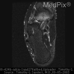
|
|
|
|
31
|
medpix__00000030
|
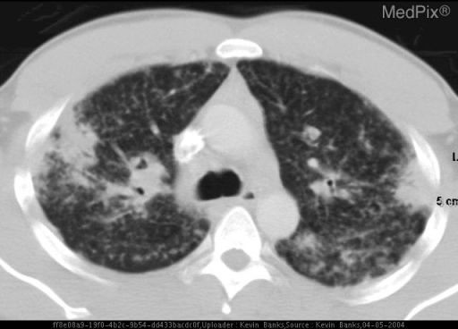
|
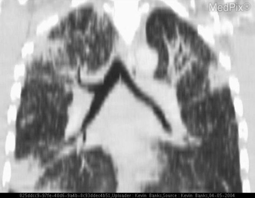
|
Title: Sarcoid
History: 36 year-old Caucasian man with worsening shortness of breath, exercise intolerance and fatigue. Referred to pulmonary for evaluation.
Findings: CXR and CT demonstrate nodular interstitial lung disease with multiple superimposed pulmonary masses/nodules. Significant mediastinal and hilar lymphadenopathy are seen. Numerous splenic and renal nodules are also present.
Diagnosis: Sarcoid
Discussion: Sarcoidosis is a multisystem granulomatous disorder of unknown etiology characterized by the presence of noncaseating granulomas in several affected organs. It is typically a disease of young adults, with a peak incidence in the third decade. The diagnosis is suggested by a combination of consistent clinical and radiological findings along with the presence of some nonspecific features such as depressed delayed-type hypersensitivity, abnormal immunoglobulins, hypercalcaemia, and elevated serum angiotensin-converting enzyme. It can be confirmed by biopsy evidence of widespread noncaseating granulomas. Pulmonary involvement accounts for most of the morbidity and mortality associated with sarcoidosis. The disease has a worse prognosis in blacks than whites, with a greater tendency for chronicity and multiorgan involvement.

About 90% of patients will have an abnormal chest radiograph at some stage, showing lymphadenopathy, parenchymal opacities, or both. Other findings are uncommon and seldom occur in isolation. Sarcoidosis is traditionally staged according to its appearance on the chest radiograph: stage I, lymphadenopathy; stage II, lymphadenopathy with parenchymal opacity; stage III, parenchymal opacity alone. Low stages at presentation carry a better prognosis than high stages.

Lymphadenopathy is evident on the chest radiograph in about 70–80% of patients at some time during the course of the condition. Sarcoidosis is characterized by bilateral, symmetrical hilar and paratracheal lymphadenopathy. In 90% of patients with lymphadenopathy, nodal enlargement is maximal on the first radiograph and usually disappears within 6–12 months. In about 5%, however, large nodes persist more or less indefinitely, and these can be a source of confusion when found later with other incidental chest pathology. Recurrence of lymphadenopathy is virtually unrecorded. The lymph nodes may calcify, sometimes in a characteristic eggshell fashion. This latter feature is shared by only a few conditions.

About 40% of patients presenting with nodal enlargement will develop parenchymal opacities, usually within a year, and of these, about one third will go on to have persistent (fibrotic) shadowing. Nodal enlargement does not develop after parenchymal opacities have appeared.

Parenchymal changes probably occur histologically in all patients but are only detected on the chest radiograph in 50–70% of cases. They may be classified as reversible, nonreversible (fibrotic), and mixed. Changes may resolve completely (two thirds of cases) or in part, leaving irreversible fibrosis. There are three major patterns of reversible opacity: irregular or rounded 2-4mm nodules, patchy airspace consolidation, or rarely 1-4cm irregular nodules. Characteristically they appear as the nodal enlargement is subsiding, in contrast to lymphoma in which these abnormalities tend to progress in unison.

Parenchymal opacities are well demonstrated on high-resolution CT. High-resolution CT is very sensitive and the findings are frequently highly specific for sarcoidosis. The principal abnormality is 1–5 mm nodules distributed in a perilymphatic fashion, predominantly along the bronchovascular bundles and subpleurally and, to a lesser extent, along interlobular septa.

Other manifestations of sarcoidosis include pleural thickening and effusions. These are unusual though and do not occur in isolation.

Cor pulmonale, mycetoma formation and pneumothorax are all recognized complications of this fibrotic stage.
[CT w/contrast (IV), Axial] Axial CT demonstrates nodular in
|
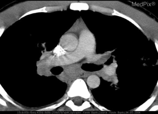
|
|
|
|
32
|
medpix__00000031
|
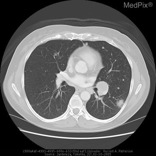
|
|
Title: Stage IV Melanoma
Migration of broken catheter
History: 52 y/o woman with hx of resected skin lesion.  Had latest PA and lateral done as part of a fever work up, with prior study in August 2004 and CT in November 2004.  Past surgical history consists of placement of a port-a-cath.
Findings: Study from December demonstrates increase in size and quantity of masses over all lung fields as compared to previous study of August in the same year.  Largest mass, measuring 3cm, is located peripherally in the left lung field.  There is prominent lymphadenopathy in both hila.  A 9.5 cm length of catheter is present in the left pulmonary artery and extends into the inferior branch.
Diagnosis: Stage IV Melanoma
Migration of broken catheter
Discussion: Discussion (include references): complication rates.  Next to skin, subcutaneous tissue, and lymph nodes, the lungs are the most common sites of melanoma to metastasize.  This is thought to be due to the capillary beds in the lung parenchyma.  Once there is metastasis to the lungs or other distant sites, the disease is stage IV and has a 5-year survival of 8-10%.  Placement of indwelling catheters is not without complications.  At the time of insertion pneumothorax is a known complication.  Once in place complications include infection, thrombosis with possible SVC syndrome, erosion of SVC, PE and breakage with migration as in this case.  Migration of the catheter has its own complications to include pulmonary infarctions, PE, perforation and arrhythmias.

Up To Date – Imagining Studies in Melanoma
Harrison’s Principals of Internal Medicine 15th ed
[CT w/contrast (IV), Axial] CT Chest with increased masses and catheter fragment in RPA.
|
|
|
|
|