| # | key | img1.png | txt |
|---|
1
|
medmax__00000000
|
|
<|img1|>
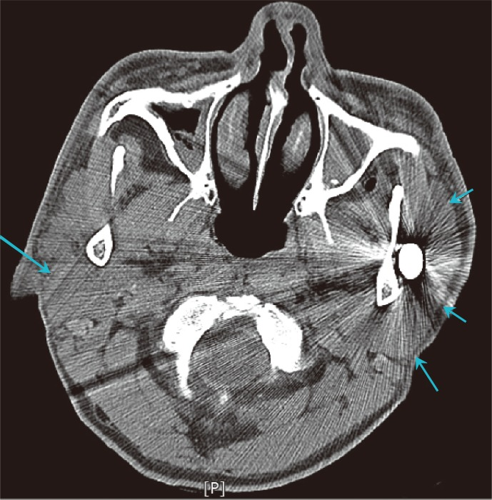
Describe the image with an emphasis on each aspect’s detail: <image><reserved08706>Facial bone computed tomography scan disclosed a radioopaque foreign body penetrating through left frontoparietal area. There was no penetration of the foreign body into left parotid gland (the blue arrows indicate both parotid glands).
|
2
|
medmax__00000001
|
|
<|img1|>
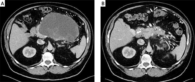
Describe the image concisely: <image><reserved08706>Contrast-enhanced abdominal computed tomography performed before and after endoscopic treatment of pancreatic necrosis. Active drainage was continued for 23 days and endoscopic necrosectomy was performed twice
|
3
|
medmax__00000002
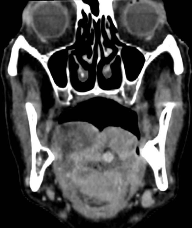
|
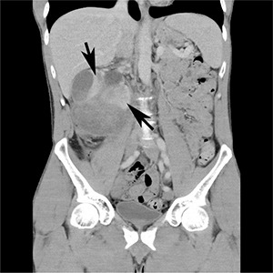
|
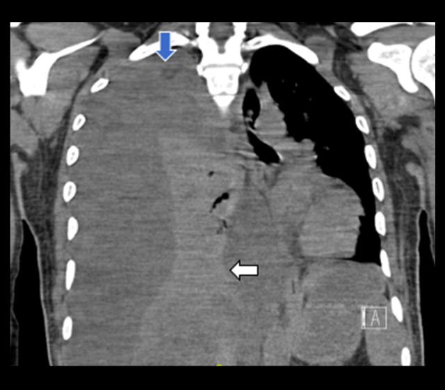
<|img1|>
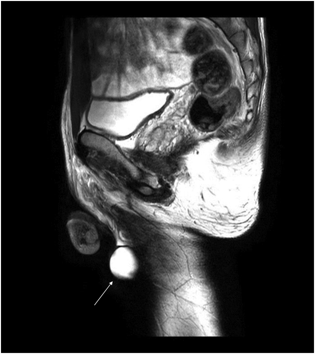
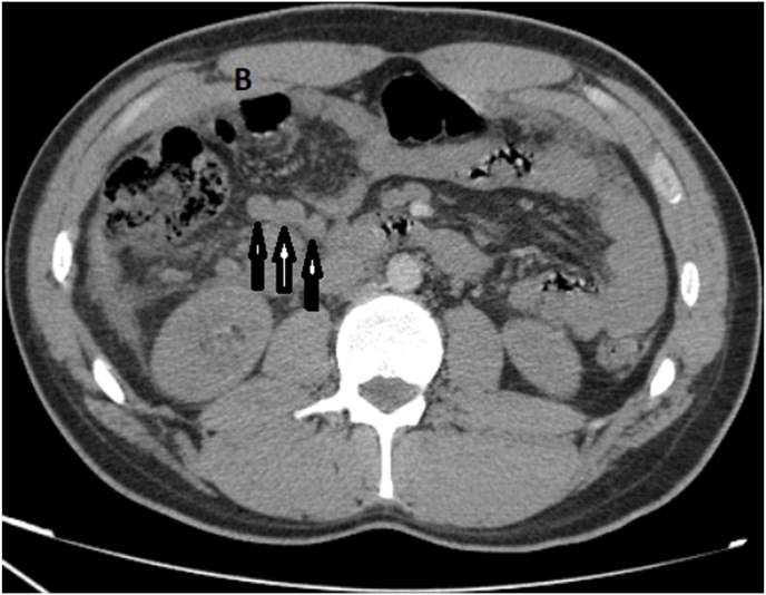
Create an image in line with the description provided: T2-weighed MRI showing a homogenous high intensity mass (white arrow) in the patient's left testicle.<reserved08706><image>
|
4
|
medmax__00000003
|
|
<|img1|>
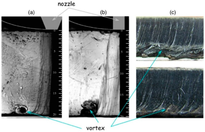
Provide a detailed description of the given image: <image><reserved08706>Vortex formed in the exit of the cut kerf using (a) converging, and (b) cylindrical nozzles. (c) Influence of their presence in the cutting quality during laser cutting of 20 mm thick mild steel using an oxygen jet (Supplying pressure p = 0.6 bar). Reprinted with permission from [37]. Copyright 2008 IOP Publishing.
|
5
|
medmax__00000004
|
|
<|img1|>
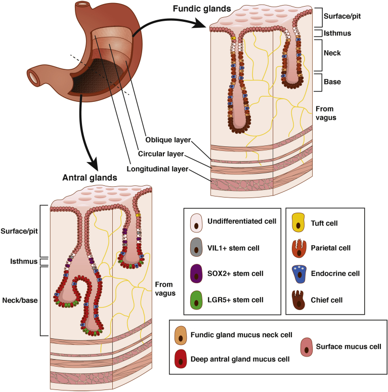
Provide a meticulous breakdown of the visual elements in the image: <image><reserved08706>Morphology and germ layer contributions of the adult stomach with specific cell types of the antral and fundic glands. The adult stomach comprises an endoderm-derived epithelial lining surrounded by mesoderm-derived smooth muscle and ectoderm-derived nerves and glia. Fundic glands are comprised of chief cells (brown) in the base, parietal (orange), endocrine (blue), and mucus (tan) cells throughout the neck, stem cells (white) in the isthmus, and mucus (pink) and tuft (yellow) cells on the surface. Antral glands comprise endocrine (blue), mucus (red), and LGR5+ (green) stem cells within the base, VIL1+ (grey), SOX2+ (purple), and other (white) stem cells in the isthmus, and mucus (pink) and tuft (yellow) cells along the surface. Myenteric neurons innervating the submucosa and muscle layers of the stomach not only stimulate hormonal secretion, including gastrin, somatostatin, and histamine, to aid in chemical digestion, but also play a role in regulating muscular contraction to aid in mechanical digestion.
|
6
|
medmax__00000005
|
|
<|img1|>
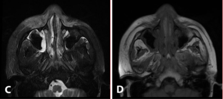
Write a precise and thorough account of the image’s contents: <image><reserved08706>continued 15-year-old male with hemophagocytic lymphohistiocytosis developed neutropenic fever and facial swelling following induction chemotherapy. T2-weighted (C) and T1-weighted (D) MRI on Day 4 demonstrate significant disease progression, with involvement of the nasal cavity and maxillary sinuses.
|
7
|
medmax__00000006
|
|
<|img1|>
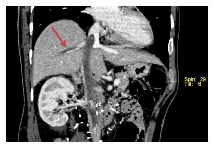
<image><reserved08706>Reformatted coronal CT image demonstrating tumour thrombus in the inferior vena cava, extending into the right hepatic vein.
|
8
|
medmax__00000007
|
|
<|img1|>
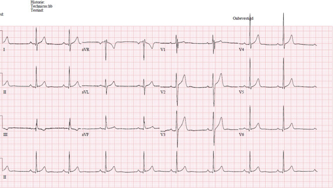
Generate an image that corresponds with Electrocardiogram with fragmented QRS complex in leads III and V1, keeping details intact.<reserved08706><image>
|
9
|
medmax__00000008
|
|
<|img1|>
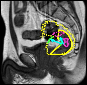
Narrate the contents of the image with precision: <image><reserved08706>Delineations of the rectal wall (yellow), nearby rectal wall (green) and tumor (purple) on a sagittal MRI scan. The dashed delineations are overlaid from a different treatment day. Note the difference in amount of displacement and direction, between tumor and nearby rectal wall (cyan arrows)
|
10
|
medmax__00000009
|
|
<|img1|>
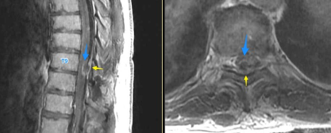
Generate a visual depiction that aligns with this description: T1 post-contrast sagittal and axial thoracic MRI showing an intramedullary enhancing lesion (blue). Epidural fluid collection (yellow) status post stimulator lead removal is also shown. MRI: magnetic resonance imaging<reserved08706><image>
|
11
|
medmax__00000010
|
|
<|img1|>
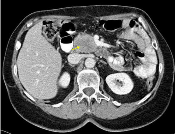
Clarify the contents of the displayed image with great detail: <image><reserved08706>Computed tomography of the abdomen four years after Billroth II resection for gastric cancer, revealing an inhomogenous mass in the pancreatic head, 4 cm in diameter. (Picture courtesy the Division of Radiology, German Cancer Research Center, provided by PD Dr. med. S. Delorme).
|
12
|
medmax__00000011
|
|
<|img1|>
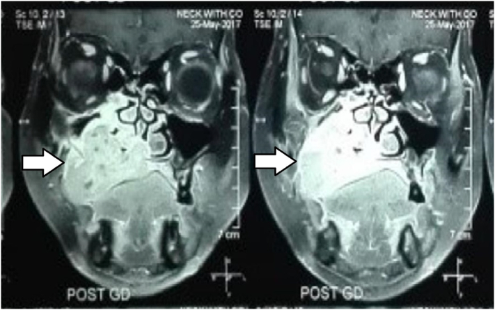
Describe the image concisely: <image><reserved08706>CT scan showing involvement of right maxillary sinus and extension to the hard palate.
|
13
|
medmax__00000012
|
|
<|img1|>
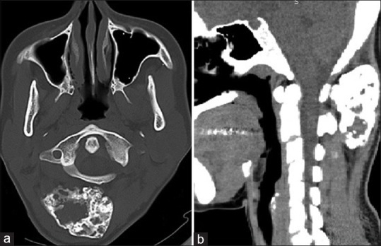
Generate an image that matches (a and b) Axial and sagittal sections of computed tomography showing enchondroma of the atlas with clarity and accuracy.<reserved08706><image>
|
14
|
medmax__00000013
|
|
<|img1|>
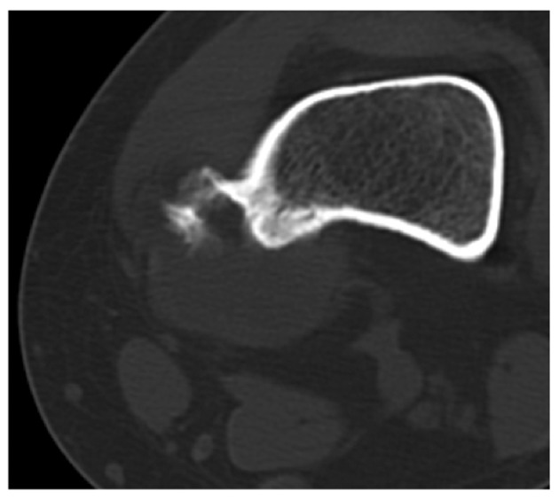
Generate a clear and precise visual based on the following text: Axial computed tomography revealing that the tumor contained calcific densities characteristic of the external bone surface and associated with a thickened cortex, which indicates a periosteal chondrogenic tumor.<reserved08706><image>
|
15
|
medmax__00000014
|
|
<|img1|>
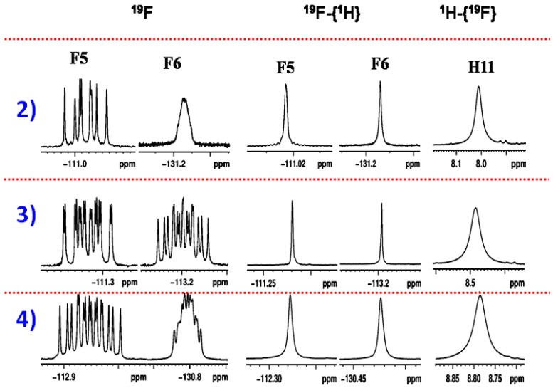
Generate an image that visually corresponds with this description: The 400 MHz 19F, 19F-{1H} and 1H-{19F} NMR spectra of molecules 2, 3 and 4 in CDCl3.<reserved08706><image>
|
16
|
medmax__00000015
|
|
<|img1|>
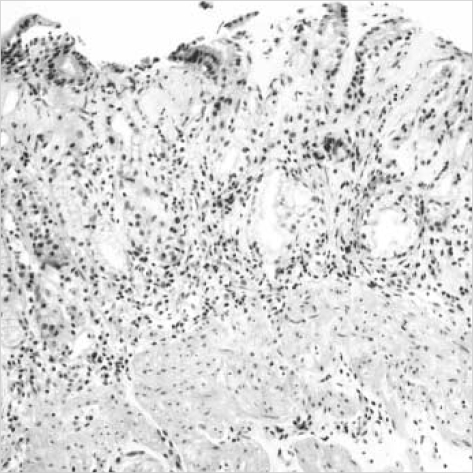
Highlight the key elements of the image briefly: <image><reserved08706>Endoscopic biopsy from the antral mucosa shows subepithelial collagenous deposition, slough of surface epithelium, inflammatory cell infiltration, and hypertrophy of muscularis mucosa (H&E, ×100).
|
17
|
medmax__00000016
|
|
<|img1|>
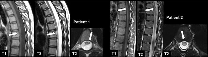
Create a detailed narrative that explains every part of the image: <image><reserved08706>MRI of a 19-year-old female patient with NMOSD (patient 1) and a 20-year-old female patient with relapsing-remitting MS (patient 2) with sagittal T1-w, sagittal T2-w, and axial T2-w images (from left to right). Note the long-segment T1-w hypointense lesion in the thoracic spinal cord on the left side of patient 1 with concomitant atrophy of the spinal cord and the focal T1-w hypointense lesion in patient 2 (arrows), compatible with black holes
|
18
|
medmax__00000017
|
|
<|img1|>
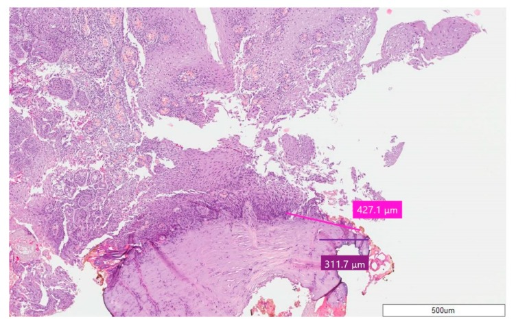
Illustrate a graphic that aligns well with Case 8—Squamous Papilloma; stain H&E, magnification 5X. The pink line for epithelium and purple line for connective tissue point out the thermal effect..<reserved08706><image>
|
19
|
medmax__00000018
|
|
<|img1|>
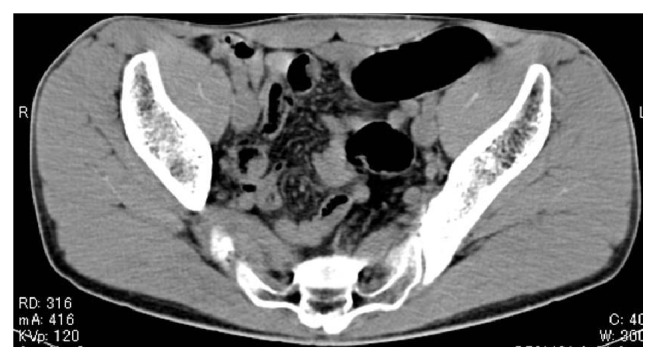
Present a quick, concise summary of the image content: <image><reserved08706>Initial computed tomography image. The image shows only normal gas distribution in the gastrointestinal tract; there is no indication of a foreign body.
|
20
|
medmax__00000019
|
|
<|img1|>
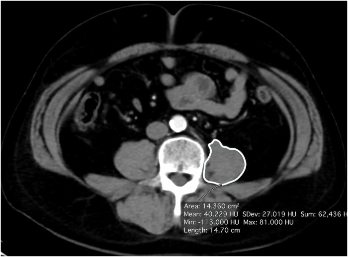
Highlight the key elements of the image briefly: <image><reserved08706>Psoas muscle area calculated by axial CT.
|
21
|
medmax__00000020
|
|
<|img1|>
Illustrate an accurate representation of the following description: Coronal venous CT image, revealing the extension of the lesion to the two posterior thirds of the tongue and an enhancing pseudoaneurysm within the necrotic/ulcerative centre of the mass.<reserved08706><image>
|
22
|
medmax__00000021
|
|
<|img1|>
Share a concise interpretation of the image provided: <image><reserved08706>CT scan – coronal view showing right kidney mass with subcapsular haematoma (arrows).
|
23
|
medmax__00000022
|
|
<|img1|>
Write a terse but informative summary of the picture: <image><reserved08706>Non-contrast-enhanced computed tomography scan of the chest illustrating massive right-sided pleural effusion (blue arrow) with complete collapse of the right lung and intraabdominal communication (white arrow).
|
24
|
medmax__00000023
|
|
<|img1|>
Present a quick, concise summary of the image content: <image><reserved08706>CT scan of abdomen (axial plane) reveal mesenteric and para-aortic lymphadenopathy (three vertical arrows).
|
25
|
medmax__00000024
|
|
<|img1|>
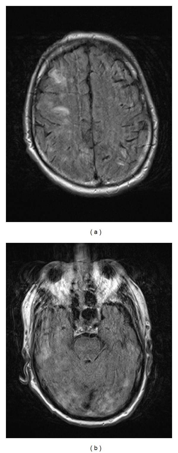
Using this text as guidance, FLAIR AXIAL MRI images (TR 9000, TE 112) from the second admission demonstrating significant interval worsening of bilateral cortical hyperintensities throughout both cerebral hemispheres. Note the mild patient motion artifact., produce an accurate image.<reserved08706><image>
|
26
|
medmax__00000025
|
|
<|img1|>
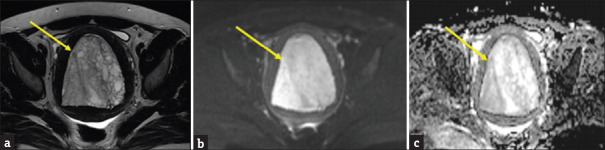
Describe the image with an emphasis on each aspect’s detail: <image><reserved08706>Endometrial hyperplasia in a 45-year-old female with: (a) Axial T2-weighted image demonstrating markedly distended endometrial cavity with smooth compressed junctional zone all around, internal morphology is smooth marginated Sol with multiple cysts of varying sizes showing predominant high T2 signal. (b) High b-value (b = 800) diffusion-weighted image shows high SI and (c) apparent diffusion coefficient map shows high SI (arrow) with values 1.7–2.1, which indicates no diffusion restriction
|
27
|
medmax__00000026
|
|
<|img1|>
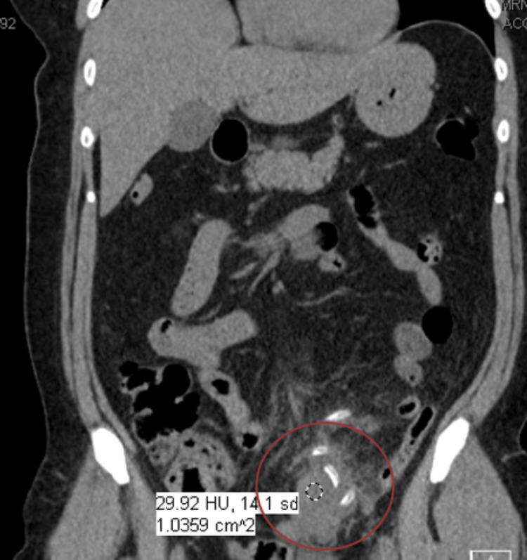
Generate an accurate representation aligned with this description: Computed tomography (CT) abdomen/pelvis without contrast showing peritoneal catheter was wrapped within the omentum and phlegmon (in red circle). <reserved08706><image>
|
28
|
medmax__00000027
|
|
<|img1|>
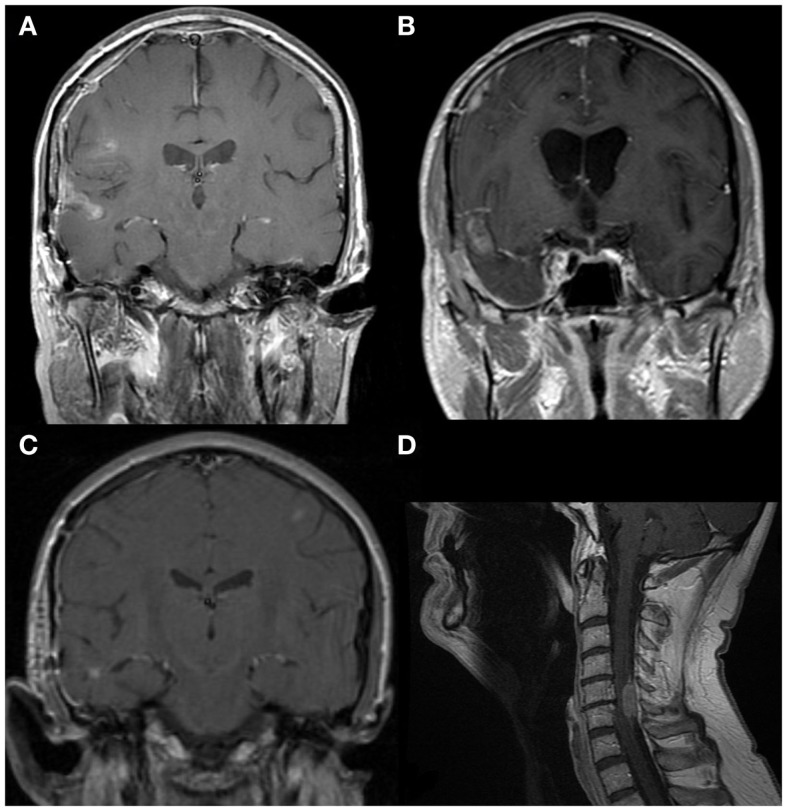
Walk through every feature visible in the image and describe it: <image><reserved08706>MRIs (1.5T, T1-weighted, post-contrast). (A) GBM with local spread to the right superior temporal sulcus. Further spread rostrally along the juxta-cortical leptomeningeal space is present. (B) GBM with local spread to the right transverse sulcus. Further spread rostrally along the juxta-cortical leptomeningeal space can also be seen. (C) A small focus of GBM in the right temporal lobe with spread to the right middle temporal sulcus, then rostrally along the subarachnoid space on the right. A second focus is seen in the left middle frontal gyrus. This is likely to have spread through the leptomeningeal space. (D) GBM with spread to dura at C5–6 and minimal intramedually invasion. Intrathecal chemotherapy is unlikely to penetrate this fully and flow caudally is likely to be impaired.
|
29
|
medmax__00000028
|
|
<|img1|>
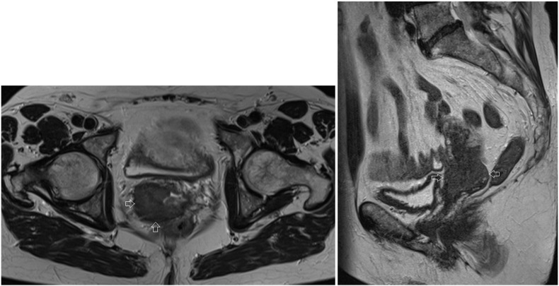
Examine each component of the image and describe it in full: <image><reserved08706>Axial and sagittal T2 MRI image of the patient before treatment. Arrows indicate the cervical tumor on the right side of the cervix, measuring 38 × 35 × 38 mm, with extension to the uterus, the right parametrium and the upper part of the vagina.
|
30
|
medmax__00000029
|
|
<|img1|>
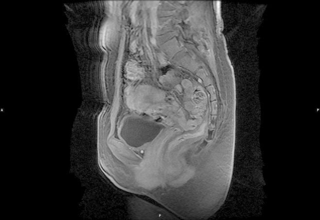
Produce a visual summary of the content in this description: MRI showed uniform enhancement after gadolinium administration on T2-weighed images.<reserved08706><image>
|
31
|
medmax__00000030
|
|
<|img1|>
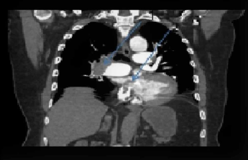
Create a thorough description covering all features of the image: <image><reserved08706>Computed tomography (CT) scan of the chest with intravenous (IV) contrast, coronal section. 1. Right main pulmonary artery occluded with thrombus. 2. A clot is clearly visible to be traversing from the right atrium to the left through the patent foramen ovale (PFO).
|
32
|
medmax__00000031
|
|
<|img1|>
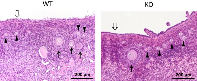
<image><reserved08706>ZP4 ablation does not cause significant alterations in folliculogenesis. Histological sections of WT and ZP4 KO ovaries stained with haematoxylin-eosin, showing an overtly normal histological structure of ZP4 KO ovaries. Arrowheads point primordial follicles, solid arrows ovarian follicles and empty arrow the germinal epithelium.
|