#692
/capstor/store/cscs/swissai/infra01/vision-datasets/raw/archive/hf___openmed-community___multicare-images
| kind | parquet |
|---|---|
| status | active |
| samples | 161,702 |
| counted via | parquet footer |
| size | 3.2 GB |
| files | 13 |
| first seen | 2026-07-22 13:56 |
| last seen | 2026-07-22 13:56 |
| registered | 2026-07-22 13:56 |
# dataset #692
path: /capstor/store/cscs/swissai/infra01/vision-datasets/raw/archive/hf___openmed-community___multicare-images
kind: parquet | status: active
samples: 161,702 (parquet footer) | size: 3.2 GB | files: 13

fields:
- file_id: text
- file: text
- main_image: text
- image_component: text
- patient_id: text
- license: text
- caption: text
- case_substring: json
- image_type: text
- image_subtype: text
- radiology_region: text
- radiology_region_granular: text
- radiology_view: text
- image: image
- labels: json
- semi_labels: json

tree (truncated):
```
.cache/huggingface/.gitignore
.cache/huggingface/download/.gitattributes.lock
.cache/huggingface/download/.gitattributes.metadata
.cache/huggingface/download/README.md.lock
.cache/huggingface/download/README.md.metadata
.cache/huggingface/download/data/train-00000-of-00002.parquet.lock
.cache/huggingface/download/data/train-00000-of-00002.parquet.metadata
.cache/huggingface/download/data/train-00001-of-00002.parquet.lock
.cache/huggingface/download/data/train-00001-of-00002.parquet.metadata
.gitattributes
README.md
data/train-00000-of-00002.parquet
data/train-00001-of-00002.parquet
```
samples
| # | file_id | file | main_image | image_component | patient_id | license | caption | case_substring | image_type | image_subtype | radiology_region | radiology_region_granular | radiology_view | image | labels | semi_labels |
|---|---|---|---|---|---|---|---|---|---|---|---|---|---|---|---|---|
1 |
file_0000000 |
PMC10000323_jbsr-107-1-3012-g3_undivided_1_1.webp |
PMC10000323_01_jbsr-107-1-3012-g3.jpg |
undivided |
PMC10000323_01 |
CC BY |
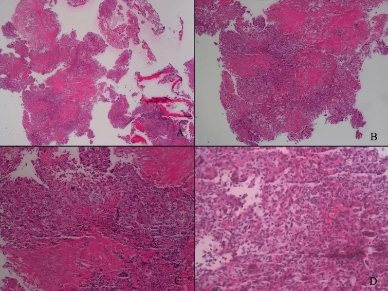
Pathological result. |
|
pathology |
h&e |
|
|
|
|||
2 |
file_0000001 |
PMC10000728_fmed-09-985235-g001_A_1_3.webp |
PMC10000728_01_fmed-09-985235-g001.jpg |
a |
PMC10000728_01 |
CC BY |
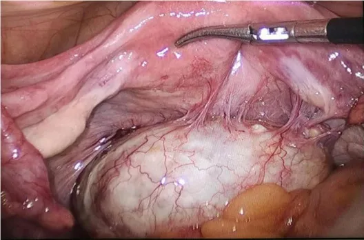
Intraoperative exploration revealed a teratoma in the utero-rectal fossa, adherent to the posterior uterine wall, right adnexa, and mesentery (A). |
|
medical_photograph |
other_medical_photograph |
|
|
|
|||
3 |
file_0000002 |
PMC10000728_fmed-09-985235-g001_B_2_3.webp |
PMC10000728_01_fmed-09-985235-g001.jpg |
b |
PMC10000728_01 |
CC BY |
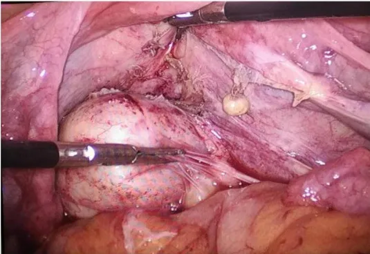
The teratoma was disconnected from the posterior uterine wall, right adnexa, and mesentery (B). |
|
medical_photograph |
other_medical_photograph |
|
|
|
|||
4 |
file_0000003 |
PMC10000728_fmed-09-985235-g001_C_3_3.webp |
PMC10000728_01_fmed-09-985235-g001.jpg |
c |
PMC10000728_01 |
CC BY |
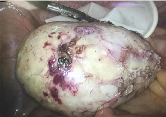
Gross observation of the specimen (C). |
|
medical_photograph |
other_medical_photograph |
|
|
|
|||
5 |
file_0000004 |
PMC10000728_fmed-09-985235-g002_A_1_3.webp |
PMC10000728_01_fmed-09-985235-g002.jpg |
a |
PMC10000728_01 |
CC BY |
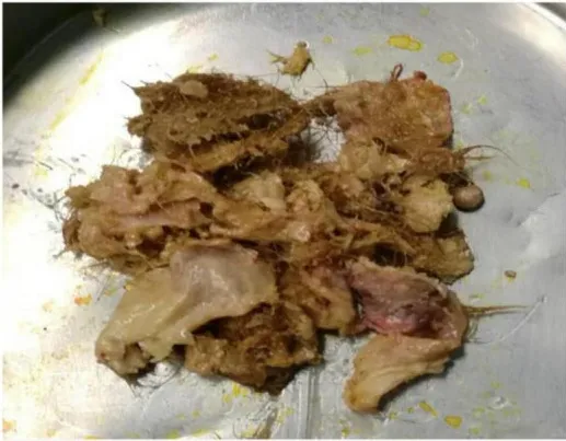
The specimen contains hair, bone, and tooth-like tissues (A). |
|
medical_photograph |
other_medical_photograph |
|
|
|
|||
6 |
file_0000005 |
PMC10000728_fmed-09-985235-g002_B_2_3.webp |
PMC10000728_01_fmed-09-985235-g002.jpg |
b |
PMC10000728_01 |
CC BY |
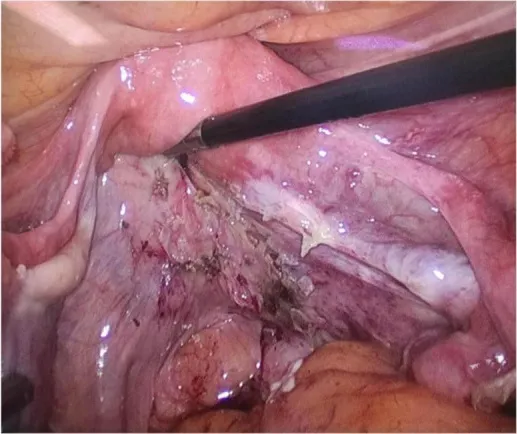
The pelvis and bilateral adnexal areas after complete resection of the teratoma showed no obvious abnormality (B). |
|
pathology |
fish |
|
|
|
|||
7 |
file_0000006 |
PMC10000728_fmed-09-985235-g002_C_3_3.webp |
PMC10000728_01_fmed-09-985235-g002.jpg |
c |
PMC10000728_01 |
CC BY |
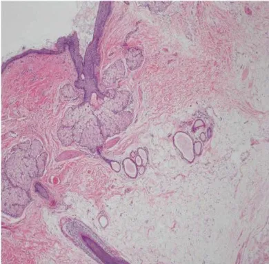
Histological pictures (C). |
|
pathology |
h&e |
|
|
|
|||
8 |
file_0000007 |
PMC10003280_JCD-26-113-g002_a_1_3.webp |
PMC10003280_04_JCD-26-113-g002.jpg |
a |
PMC10003280_04 |
CC BY-NC-SA |
Medical image. |
|
pathology |
giemsa |
|
|
|
|||
9 |
file_0000008 |
PMC10003280_JCD-26-113-g002_b_2_3.webp |
PMC10003280_04_JCD-26-113-g002.jpg |
b |
PMC10003280_04 |
CC BY-NC-SA |
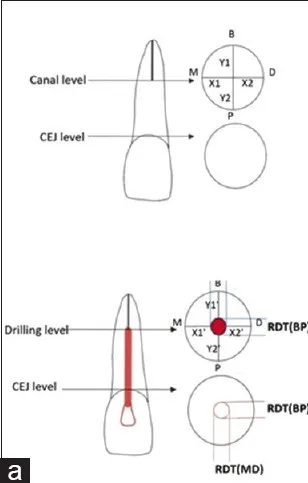
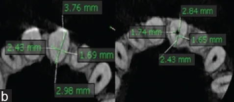
Schematic view of calculation of linear deviation. |
|
radiology |
ct |
thorax |
thorax |
sagittal |
|
|
|
10 |
file_0000009 |
PMC10003280_JCD-26-113-g002_c_3_3.webp |
PMC10003280_04_JCD-26-113-g002.jpg |
c |
PMC10003280_04 |
CC BY-NC-SA |
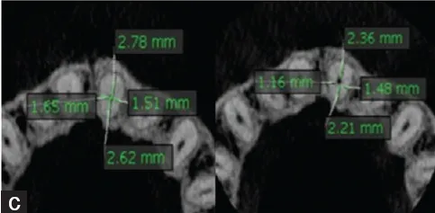
Dentin thickness at CEJ level. Dentin thickness at canal level. CEJ: Cementoenamel junction. |
|
radiology |
mri |
thorax |
thorax |
ultrasound_view |
|
|
|