| # | key | img1.png | txt |
|---|
1
|
medtrinity_conv_1__00000000
|
|
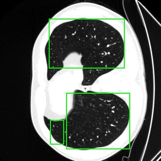
user: Provide a concise description of this image, focusing on the main subject and background. <|img1|>
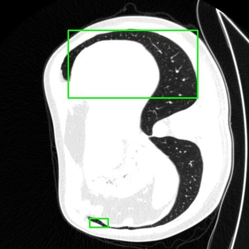
assistant:  The image is a transverse thoracic CT scan, displaying the lungs, heart, and part of the spine.

- The **lungs** are prominently visible, with the **heart** centrally positioned between them.
- A region of interest is located in the **right-center and lower-middle** of the image, covering approximately **13.6%** of the total area.
- This region shows an **abnormal texture and density** compared to the surrounding healthy lung tissue, indicating a possible pathological condition.
- The abnormality appears closely related to adjacent lung tissue, suggesting it may be:
  - A direct site of disease,
  - Or an area jointly affected by an underlying process.
- Based on available information, this finding could represent:
  - **Non-enhancing pneumonia**, or
  - A **mediastinal structure** with associated lung pathology.
- The spatial relationship between this abnormality and surrounding regions may reflect the disease’s impact on lung structure and function.
|
2
|
medtrinity_conv_1__00000001
|
|
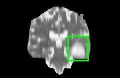
user: Provide a concise description of this image, focusing on the main subject and background. <|img1|>
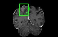
assistant:  The image is a magnetic resonance imaging (MRI) scan of the brain, displaying varying shades of gray that correspond to different brain tissues and structures.

A region of interest is located in the right-center horizontally and lower-middle vertically, covering approximately 5.2% of the total image area. This region exhibits an abnormal appearance compared to the surrounding brain tissue, with distinct texture and contrast differences that may indicate a pathological process—potentially a brain tumor.

Key observations:
- The abnormality stands out due to its unique visual characteristics relative to adjacent brain matter.
- Its presence may suggest compression or displacement of nearby brain structures, which is commonly associated with tumor growth.
- The exact nature of the relationship between the abnormality and surrounding tissue depends on the tumor’s type and growth pattern.

Further clinical evaluation is recommended to confirm the diagnosis.
|
3
|
medtrinity_conv_1__00000002
|
|
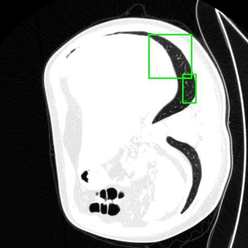
user: Provide a concise description of this image, focusing on the main subject and background. <|img1|>
assistant:  The image is a high-resolution computed tomography (HRCT) scan of the chest, capturing the thoracic cavity with clear visualization of the lungs.

Key anatomical structures visible:
- Right and left lungs
- Heart shadow in the central region
- Portions of the rib cage

The region of interest is located in the right-center and upper-middle portion of the scan. Here, an area of abnormal lung tissue is evident, characterized by:

- Altered texture and density
- Findings consistent with **ground-glass opacities** or **consolidation**

These imaging features are hallmark signs of **COVID-19 infection**, as supported by established medical knowledge. The abnormality is localized and bordered by otherwise normal lung tissue, suggesting a focal infectious process likely resulting from direct viral invasion of the lung parenchyma, leading to localized inflammation.
|
4
|
medtrinity_conv_1__00000003
|
|
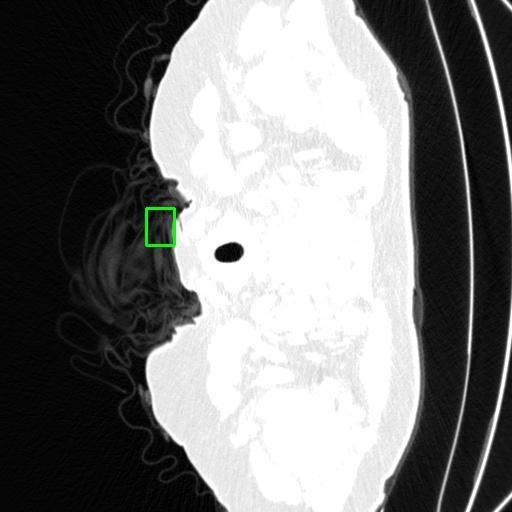
user: Create an accurate image description for a machine learning dataset, covering all key elements. <|img1|>
assistant:  The image is a high-resolution computed tomography (HRCT) scan of the lungs, showing the thoracic cavity with the heart centered and the lungs on either side.

A region of interest, located in the left-center horizontally and the middle vertically, occupies approximately 0.4% of the total area. This region exhibits an unusual texture and density, consistent with findings typically associated with COVID-19 infection.

Key observations:
- The abnormality is situated within the lung parenchyma, a common site affected by COVID-19.
- The appearance aligns with characteristic features of COVID-19 pneumonia, including ground-glass opacities and consolidation.
- These changes reflect altered lung tissue density and texture, often resulting from inflammation and infection spread.

The findings suggest a possible progression of the disease, with potential for involvement of adjacent lung areas due to the inflammatory response.
|
5
|
medtrinity_conv_1__00000004
|
|
user: What do you see in this image? <|img1|> Describe it in detail.
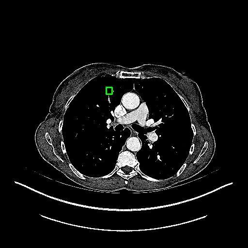
assistant:  The chest CT scan shows a transverse section of the thorax, highlighting the lungs, heart, and posterior spine. The lungs are the dominant structures, with the heart centrally positioned between them and the spine visible at the back.

A region of interest is identified in the upper-middle, horizontally centered portion of the image. This area exhibits a distinct texture or density compared to the surrounding lung tissue, suggesting a possible lesion. The abnormality is located within the lung parenchyma and does not appear to involve the heart, spine, or other major structures.

The observed change may represent a pathological condition such as:
- Ground-glass opacity
- Consolidation
- Or another form of lung parenchymal abnormality

This lesion could indicate a localized lung pathology, potentially reflecting either a focal process or a manifestation of a broader disease. Its presence may affect the function or integrity of adjacent lung tissue, warranting further clinical evaluation.
|
6
|
medtrinity_conv_1__00000005
|
|
user: Create an image caption that would help someone understand the picture without seeing it. <|img1|>
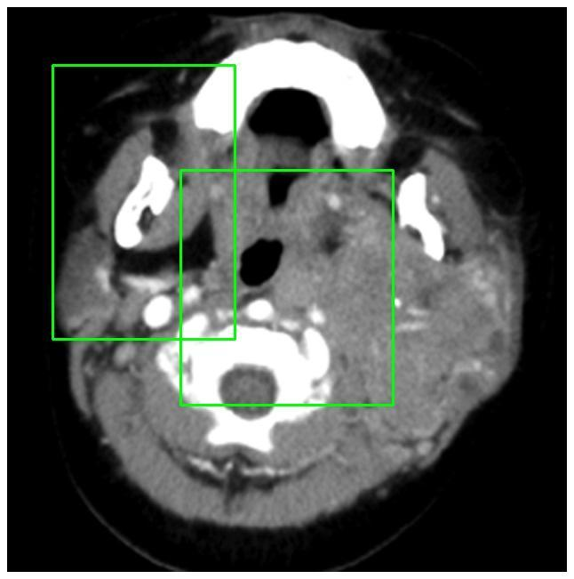
assistant:  The image is a transverse CT scan of the abdomen, but the focus is on the neck region.

### Key Details:
- **Region of Interest (ROI)**:
  Located in the left-center horizontally and upper-middle vertically.
  Occupies approximately **15.0%** of the total image area.

- **Visible Structures**:
  - Cervical vertebrae
  - Upper portions of the lungs

### Abnormality in the Region of Interest:
- The ROI shows an **abnormal texture and density** compared to surrounding tissues.
- This suggests a **potential pathology** involving the cervical spine and/or adjacent soft tissues.

### Clinical Implications:
- Due to its **central location in the neck**, this abnormality may:
  - Affect nearby critical structures (e.g., airway, blood vessels, nerves).
  - Be influenced by or spread to adjacent tissues.
- The close anatomical relationships in the neck mean this finding could indicate:
  - A primary disease process in the cervical region.
  - Secondary involvement from nearby organs or systemic conditions.

Further evaluation is warranted to determine the nature and clinical significance of this finding.
|
7
|
medtrinity_conv_1__00000006
|
|
user: Generate a caption that identifies any visible text, signs, or labels in the image. <|img1|>
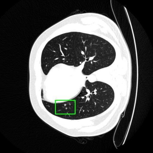
assistant:  The image is a high-resolution computed tomography (HRCT) scan of the chest, displaying the lungs, heart, and part of the liver.

- **Primary structures visible**:
  - The lungs are the dominant feature.
  - The heart is centrally located between the lungs.

- **Region of interest**:
  - Located in the lower-middle horizontal center of the image.
  - Shows a localized area of abnormal tissue density within the lung.

- **Possible interpretation**:
  - The abnormality appears as a change consistent with **ground-glass opacities** or **consolidation**, which are hallmark findings in **COVID-19 infection**.

- **Context and implications**:
  - The abnormality is surrounded by normal lung parenchyma, suggesting the condition may be **localized** or in an **early stage**.
  - Its proximity to healthy tissue raises the possibility of potential spread, though the image alone cannot confirm directionality or progression.

> Note: This finding is suggestive but not diagnostic; clinical correlation and additional testing are required.
|
8
|
medtrinity_conv_1__00000007
|
|
user: Provide an objective description of what appears in the image. <|img1|>
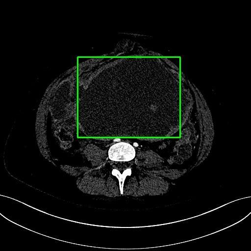
assistant:  The image is a transverse CT scan of the abdomen, focusing on the upper-middle region. It displays key abdominal structures, including:

- **The liver**: The largest organ, located in the upper right quadrant.
- **The spine and ribs**: Visible as dense, bony structures.
- **Parts of the gastrointestinal tract**: Likely present but not clearly delineated.

A distinct area of interest is centrally located in the upper-middle portion of the scan. This region exhibits an unusual texture and density compared to surrounding tissues, suggesting a possible lesion. Potential causes include:

- A mass or tumor
- A cyst
- Other pathological changes

The nature of this lesion may involve interactions with adjacent organs or tissues:

- If it is a **tumor**, it could exert pressure on or infiltrate nearby structures.
- If it is due to an **infectious process**, it may have originated from or spread to adjacent organs.

The exact implications depend on the underlying pathology, which requires further clinical evaluation. Overall, the scan highlights an abnormality in the upper-middle abdomen that warrants careful assessment in context with the surrounding anatomy.
|
9
|
medtrinity_conv_1__00000008
|
|
user: Create an accurate image description for a machine learning dataset, covering all key elements. <|img1|>
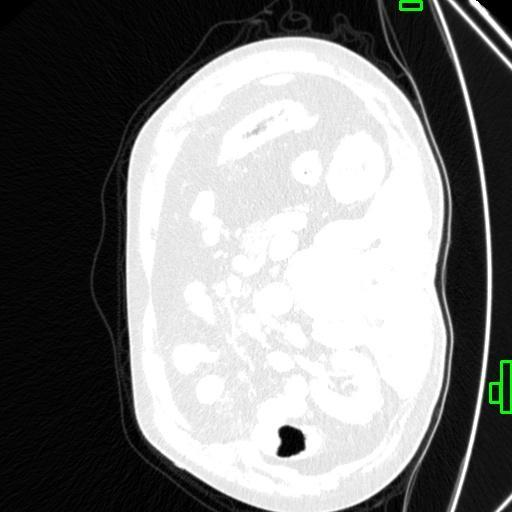
assistant:  The image is a transverse CT scan of the abdomen, highlighting key organs including the liver, which dominates the upper right quadrant.

A region of interest is identified in the lower-middle right portion of the image. This area corresponds to the lower lobe of the right lung and displays a small region of altered density, suggesting a potential abnormality.

Key observations:
- The abnormality is distinct from the surrounding lung tissue.
- It is located adjacent to the liver, indicating close anatomical proximity.
- This proximity may imply a possible relationship in terms of shared blood supply or mechanical influence.

While the finding could indicate a localized lung condition, its location near the liver warrants consideration of potential interactions between the lung and liver tissues. Further clinical evaluation is recommended to determine the nature and significance of this abnormality.
|
10
|
medtrinity_conv_1__00000009
|
|
user: Generate a caption that accurately represents the content of this image. <|img1|>
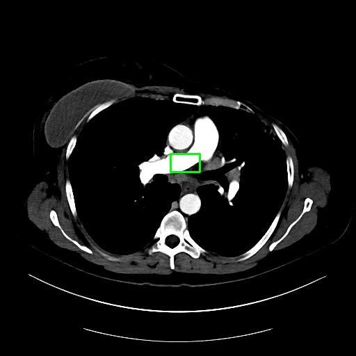
assistant:  The image is a transverse CT scan of the chest, displaying the lungs, heart, and part of the spine.

- The **heart** is centrally positioned, with the **lungs** on either side and the **spine** visible at the back.
- A region of interest is located centrally and horizontally in the middle of the image, encompassing a small portion of the heart.
- This area exhibits a distinct texture or density compared to the surrounding cardiac tissue, which may indicate a **lesion**.
- Given its proximity to the heart, this abnormality could be:
  - Influencing nearby cardiac structures, or
  - Being affected by them.
- Such a finding may suggest a **localized cardiac condition** with potential implications for cardiac function or adjacent anatomy.
|
11
|
medtrinity_conv_1__00000010
|
|
user: Describe everything visible in this image in a clear, factual way. <|img1|>
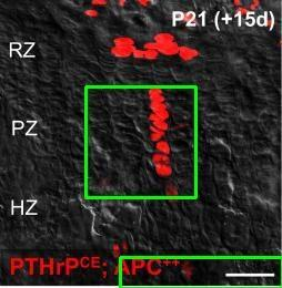
assistant:  The image is a magnetic resonance imaging (MRI) scan of the abdominal region, showing a cross-sectional view of internal structures.

- **Region of Interest**:
  Located in the lower right-center horizontally and centered vertically, this area occupies approximately 15.0% of the image. It is situated in the lower abdominal region, likely involving the pelvic area.

- **Findings**:
  The region exhibits abnormal tissue characteristics, including altered signal intensity. These changes may indicate a pathological process.

- **Possible Implications**:
  The unusual features—such as differences in color, texture, or size—could reflect disease affecting pelvic tissues. Given the anatomical proximity and interconnected nature of pelvic structures, this abnormality may either originate in the region or impact surrounding tissues.

Further clinical correlation is recommended to determine the exact nature of the finding.
|
12
|
medtrinity_conv_1__00000011
|
|
user: What do you see in this image? <|img1|> Describe it in detail.
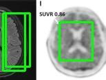
assistant:  The image is a magnetic resonance imaging (MRI) scan of the brain, providing a detailed view of its internal structures.

- A region of interest is visible on the left side of the brain, accounting for approximately 15% of the scanned area.
- This region displays an abnormality consistent with **demyelination**, characterized by:
  - Altered texture
  - Different signal intensity compared to surrounding healthy tissue
- These features are indicative of a **lesion**.
- Due to its proximity to adjacent brain structures, this lesion may interact with or affect nearby neural pathways, potentially influencing or being influenced by surrounding neural function.
|
13
|
medtrinity_conv_1__00000012
|
|
user: Create an image caption that would help someone understand the picture without seeing it. <|img1|>
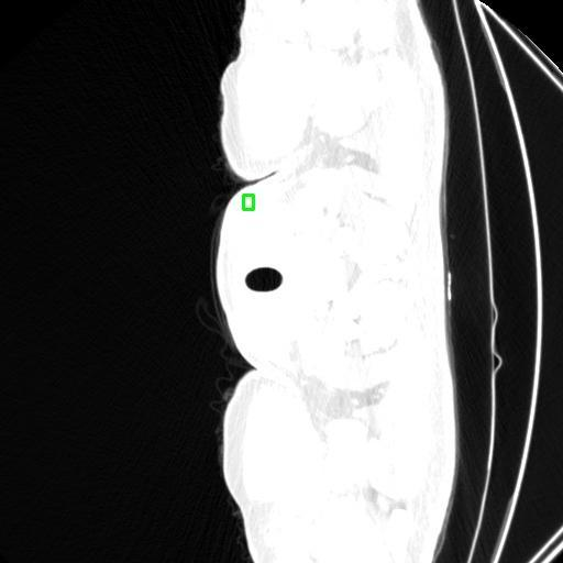
assistant:  The image is a transverse computed tomography (CT) scan of the chest, displaying the thoracic cavity, including the lungs, spine, and surrounding structures.

A region of interest is located in the **upper central portion** of the image, approximately within the **upper lobe of the right lung**. This area occupies a very small fraction of the total image (approximately 0.0%).

Key observations:
- The tissue in this region exhibits an **abnormal texture or density** compared to the surrounding lung parenchyma.
- This discrepancy may indicate a **pathological condition**, such as a mass, lesion, or localized disease process.
- The abnormality could be **interacting with adjacent lung tissue**, potentially causing a localized volume increase or reflecting a response to an underlying pathology.

Further clinical or radiological evaluation is recommended to determine the nature of this finding.
|
14
|
medtrinity_conv_1__00000013
|
|
user: Generate a caption that identifies any visible text, signs, or labels in the image. <|img1|>
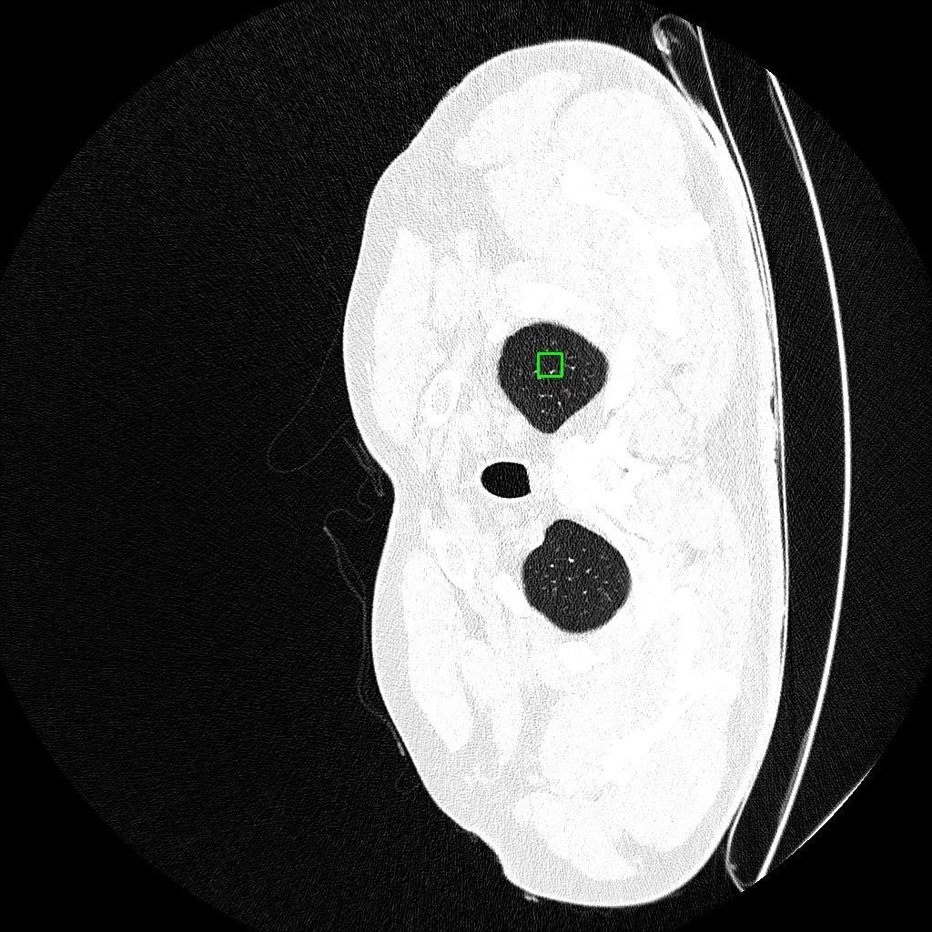
assistant:  The image is a radiograph of the thoracic and upper abdominal regions, showing the lungs, heart, ribs, and liver. No medical devices are visible.

- **Lungs**: Located in the upper portion of the image.
- **Heart and mediastinal structures**: Centered in the image.
- **Liver**: Visible in the lower portion.

A green bounding box is positioned in the upper-middle area of the image, highlighting an abnormality in the **right first rib**. The rib exhibits a **hypoplasmic appearance**—it is smaller than normal and fails to articulate properly with the manubrium.

This finding may be associated with **congenital pseudoarthrosis of the left first rib**. Both ribs are part of the rib cage and could be affected by similar congenital developmental anomalies.
|
15
|
medtrinity_conv_1__00000014
|
|
user: Provide a concise description of this image, focusing on the main subject and background. <|img1|>
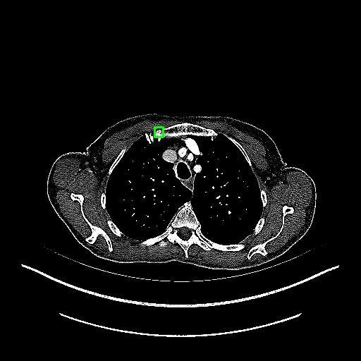
assistant:  The image is a transverse CT scan of the chest, displaying the lungs, heart, and part of the spine.

- **Lungs**: The primary structures visible, occupying most of the field of view.
- **Heart**: Centrally located between the lungs.
- **Spine**: Visible posteriorly.

A region of interest is identified in the **upper-middle, horizontally centered** portion of the image. This area exhibits an **abnormal texture or density** compared to the surrounding lung tissue, suggesting the presence of a **possible lesion**.

Key observations:
- The abnormality is located within the lung parenchyma.
- Its proximity to adjacent lung regions suggests it may be related to or influenced by surrounding tissue.
- This finding could indicate either:
  - A **primary lung condition**, such as infection, inflammation, or neoplasm.
  - A **secondary manifestation** of a systemic disease.

Further clinical correlation and imaging follow-up are recommended to determine the nature of this abnormality.
|
16
|
medtrinity_conv_1__00000015
|
|
user: Provide an objective description of what appears in the image. <|img1|>
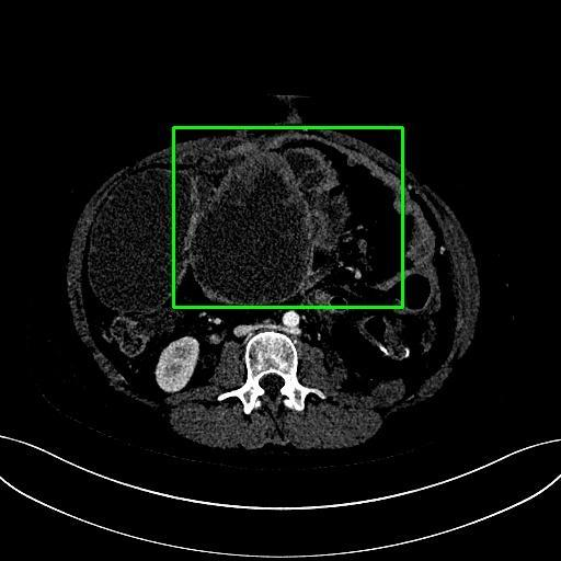
assistant:  The image is a transverse CT scan of the abdomen, centered on the upper-middle region, highlighting the liver and portions of the gastrointestinal tract.

- A region of interest, covering approximately **13.1% of the area**, displays an abnormality within the liver.
- This abnormality is visually distinct from the surrounding hepatic tissue, showing differences in **texture** and possibly **size**.
- These characteristics suggest a **pathological condition**, though the exact nature remains undetermined without further clinical evaluation.
- The liver’s close anatomical relationship with adjacent structures, including the gastrointestinal tract, may imply potential interactions between the abnormality and these organs.
- However, the precise clinical implications require additional diagnostic correlation.
|
17
|
medtrinity_conv_1__00000016
|
|
user: Explain what is shown in the image. <|img1|>
assistant:  The image is a high-resolution computed tomography (HRCT) scan of the chest, providing a cross-sectional view of the thoracic cavity. Key structures visible include:

- **Lungs**: The primary focus of the scan
- **Heart**: Located centrally, partially visible
- **Rib cage**: Surrounding the thoracic cavity

The region of interest is centrally located in the upper-middle portion of the image and shows an abnormality in the lung tissue. This abnormality is characterized by:

- Altered texture and density compared to surrounding healthy lung parenchyma
- Features consistent with **ground-glass opacities** and/or **consolidation**

These findings are commonly associated with **COVID-19 pneumonia**. The involvement of multiple lung regions suggests a diffuse pattern, which is typical of COVID-19, as the disease often affects multiple lobes and can evolve over time—either progressing or resolving as the condition advances.
|
18
|
medtrinity_conv_1__00000017
|
|
user: Create an accurate image description for a machine learning dataset, covering all key elements. <|img1|>
assistant:  The image is a magnetic resonance imaging (MRI) scan of the brain, providing a detailed view of its internal structures.

A region of interest is visible in the central, upper-middle portion of the brain. This area accounts for approximately 4.8% of the total image and is suggestive of a brain tumor.

Key characteristics of this region include:
- An abnormal appearance compared to surrounding brain tissue, potentially manifesting as differences in color, texture, or size.
- Possible effects on adjacent brain structures, such as:
  - Direct expansion causing compression or displacement of nearby tissues.
  - Secondary effects like edema (swelling) or disruption of normal brain function.

These features collectively indicate the presence of a pathological lesion consistent with a tumor.
|
19
|
medtrinity_conv_1__00000018
|
|
user: Describe everything visible in this image in a clear, factual way. <|img1|>
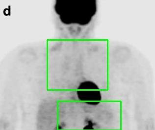
assistant:  The image is a PET-CT scan showing a cross-sectional view of the chest area, highlighting metabolic activity through the distribution of a radiotracer, typically (18)F-fluorodeoxyglucose (FDG).

Key observations:
- The scan visualizes metabolic activity in tissues and organs, including the lungs, liver, and parts of the gastrointestinal tract.
- A region of interest, located centrally and toward the bottom of the image, covers approximately 9.1% of the area.
- This region shows **unusual radiotracer uptake**, suggesting elevated metabolic activity—commonly associated with conditions such as inflammation or tumor formation.

Additional notes:
- The region is visually distinct from surrounding tissues due to its texture and intensity.
- Its exact anatomical relationship to nearby structures is not specified.
- The abnormal uptake may indicate:
  - A **primary lesion** affecting local tissues, or
  - A **secondary manifestation** of a systemic condition, potentially interacting with adjacent organs.

Further clinical correlation is recommended to determine the nature of this finding.
|
20
|
medtrinity_conv_1__00000019
|
|
user: Summarize the content of the image as if it were an alternative text (alt text) for accessibility. <|img1|>
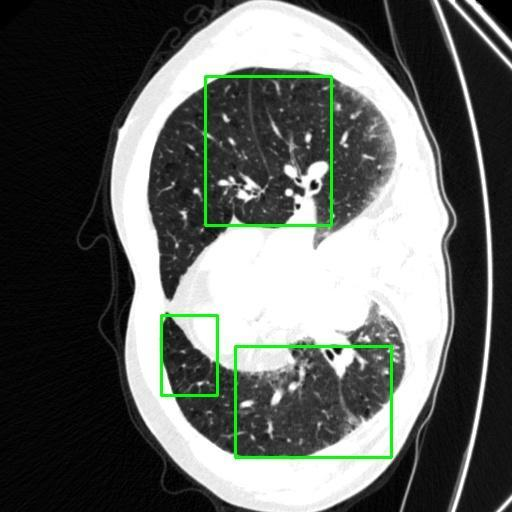
assistant:  The image is a coronal CT scan of the chest, displaying the lungs, heart, and part of the liver.

### Key Structures Visible:
- **Lungs**: The primary focus of the image, occupying most of the thoracic cavity.
- **Heart**: Centrally located between the two lungs.
- **Liver**: Visible in the lower portion of the image, beneath the right lung.

### Region of Interest:
- Located in the **right-center to lower-middle** of the image, within the **lower lobe of the right lung**.
- Exhibits an abnormal texture and density compared to the surrounding lung tissue.
- This finding is consistent with pathological changes such as:
  - **Ground-glass opacities**
  - **Consolidation**

These features suggest a potential lung disease affecting the lung parenchyma and possibly impairing normal respiratory function.

### Clinical Considerations:
- The abnormality is adjacent to the liver, but the image does not provide sufficient detail to determine whether there is direct involvement of the liver or other adjacent structures.
- The lesion may represent a localized finding or could be part of a more widespread pulmonary condition.

Further clinical correlation and additional imaging may be needed to determine the exact nature and extent of the abnormality.
|
21
|
medtrinity_conv_1__00000020
|
|
user: Provide a concise description of this image, focusing on the main subject and background. <|img1|>
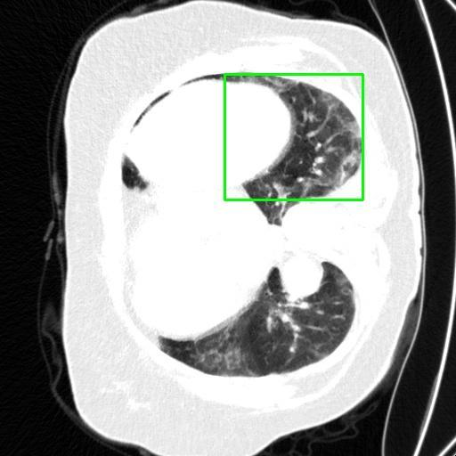
assistant:  This is a high-resolution computed tomography (HRCT) image of the lungs, showing a cross-sectional view of the thoracic cavity. Key structures visible include:

- **Lung parenchyma**
- **Heart shadow** in the mediastinum
- **Parts of the rib cage**

A region of interest is identified in the **right-center horizontally** and **upper-middle vertically**, occupying approximately **8.3% of the image area**. This region is located within the lung tissue and exhibits the following abnormalities:

- **Ground-glass opacities**
- **Consolidation**
- Other textural changes consistent with **COVID-19 pneumonia**

These findings align with known radiological features of COVID-19, which typically affect the lung parenchyma. The pattern of abnormalities suggests possible **diffuse or multifocal lung involvement**, which may reflect disease progression or response to treatment. The region of interest is likely part of a broader pattern of pulmonary changes associated with the infection.
|
22
|
medtrinity_conv_1__00000021
|
|
user: Explain what is shown in the image. <|img1|>
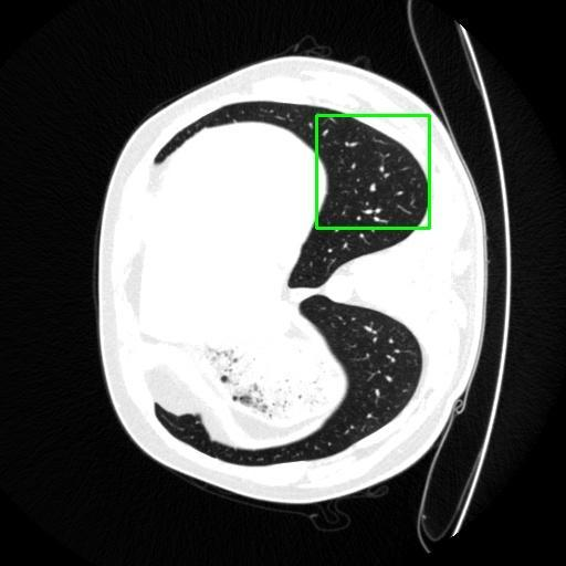
assistant:  This is a high-resolution computed tomography (HRCT) image of the lungs, showing a cross-sectional view with the heart and both lungs visible.

- **Region of Interest**:
  Located in the right-center horizontally and upper-middle vertically, this area occupies approximately 4.0% of the total image.

- **Abnormality Detected**:
  A ground-glass opacity is present within this region. This finding is characteristic of COVID-19 infection and is distinctly different from the surrounding lung tissue, which appears darker and is likely normal.

- **Anatomical Context**:
  The abnormality is situated near the heart and adjacent lung structures, raising the possibility that the disease may affect these nearby tissues due to its location.

- **Clinical Correlation**:
  The presence of ground-glass opacity in the upper lobe of the right lung aligns with the typical radiological pattern seen in COVID-19, which commonly involves ground-glass opacities and, in some cases, consolidation.
|
23
|
medtrinity_conv_1__00000022
|
|
user: Generate a caption that identifies any visible text, signs, or labels in the image. <|img1|>
assistant:  The image is a magnetic resonance imaging (MRI) scan of bone, displaying a cross-sectional view with varying shades of gray that reflect differences in tissue density.

A region of interest is centrally located toward the bottom of the image, covering approximately 6.1% of the total area. This region exhibits an abnormality characterized by:

- A distinct texture compared to surrounding bone tissue
- A noticeable difference in signal intensity

These features suggest a possible pathological process, such as:

- Ischemia (reduced blood flow leading to tissue damage)
- A benign or malignant tumor

The abnormality’s location and relationship to adjacent bone structures may indicate:

- A localized bone disorder
- Potential impact on the function of nearby structures
- Possible spread of the underlying disease process

Further clinical evaluation is recommended to determine the exact nature of the abnormality.
|
24
|
medtrinity_conv_1__00000023
|
|
user: Provide a concise description of this image, focusing on the main subject and background. <|img1|>
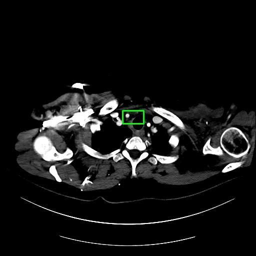
assistant:  The image is a computed tomography (CT) scan of the abdomen, showing a cross-sectional view of the spine and surrounding tissues.

- The region of interest is centrally located in the image and exhibits an abnormal texture or density compared to the surrounding tissue.
- This abnormality may indicate the presence of a lesion.
- The lesion is situated near the spine, suggesting a possible anatomical relationship with spinal structures.
- Due to its proximity, the lesion could be affecting—or being affected by—the spinal tissues.
|
25
|
medtrinity_conv_1__00000024
|
|
user: What do you see in this image? <|img1|> Describe it in detail.
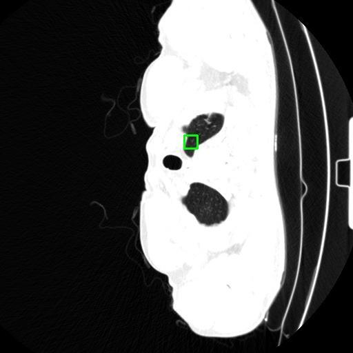
assistant:  The image is a computed tomography (CT) scan of the chest, providing a cross-sectional view of the thoracic cavity.

### Key Observations:
- **Region of Interest**:
  Located at the center of the image horizontally and midway vertically, this area occupies approximately **0.1%** of the total image.
  It is situated within the **right lung** and displays an **unusual texture and density**, consistent with findings suggestive of **COVID-19 infection**.

- **Visible Anatomy**:
  - **Right lung**: Dominates the image.
  - **Spine**: Partially visible in the background.
  - **Heart**: Upper portion may be visible, though not the primary focus.

- **Pattern of Abnormality**:
  The affected region is surrounded by **normal lung tissue**, indicating that the infection may be **localized**.
  Its proximity to adjacent lung areas suggests potential **local spread** or influence on nearby tissue, which could contribute to disease progression.

This finding supports the possibility of early or focal COVID-19 pneumonia, with no evidence yet of widespread involvement.
|
26
|
medtrinity_conv_1__00000025
|
|
user: Explain what is shown in the image. <|img1|>
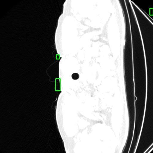
assistant:  The image is a non-contrast computed tomography (CT) scan of the thoracic region, displaying the cerebral hemispheres, ventricular system, and intrathoracic structures.

### Key Findings:
- A small, localized abnormality is visible in the **left-center at the top** of the image, occupying approximately **0.2% of the area**.
- This region is located within the **lung tissue**, adjacent to the **lateral ventricles**.
- It exhibits an unusual **texture or density difference** compared to the surrounding lung parenchyma.

### Interpretation:
- Given the context of thoracic CT imaging, this abnormality may represent a **localized lung pathology**, such as:
  - A nodule
  - A lesion
- Potential implications include:
  - Direct extension into adjacent tissue
  - Mass effect
  - Secondary changes such as edema

This finding warrants further clinical evaluation, as it could indicate an underlying pulmonary condition.
|
27
|
medtrinity_conv_1__00000026
|
|
user: Describe everything visible in this image in a clear, factual way. <|img1|>
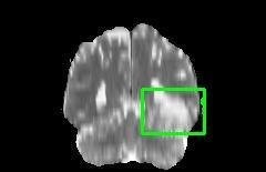
assistant:  The image is a magnetic resonance imaging (MRI) scan of the brain, revealing its detailed internal structures.

A region of interest is located in the right-center horizontally and lower-middle vertically, occupying approximately 6.0% of the total area. This region exhibits:

- An abnormal texture
- Possibly altered signal intensity

These characteristics suggest a potential brain tumor. The abnormality is embedded within the brain tissue and stands out distinctly from the surrounding healthy structures, indicating a pathological change.

The relationship between this region and adjacent brain tissue may reflect the tumor’s influence, potentially causing:

- Compression of nearby structures
- Displacement of surrounding brain tissue

These secondary effects are consistent with the presence of a mass lesion.
|
28
|
medtrinity_conv_1__00000027
|
|
user: Create an accurate image description for a machine learning dataset, covering all key elements. <|img1|>
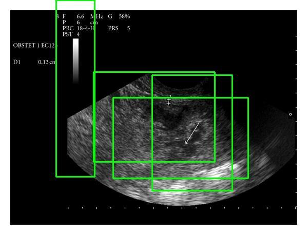
assistant:  The image is a transverse MRI section of the abdomen, but it primarily displays cerebral structures.

### Key Observations:
- **Region of Interest (ROI)**:
  Located in the **upper-middle right-center** of the image, occupying approximately **15.0%** of the total area.

- **Visible Anatomy**:
  - Cerebral hemispheres are clearly visible.
  - The cerebellum is present in the lower portion of the image.

- **Abnormal Findings**:
  The ROI exhibits **abnormal signal intensity**, with:
  - A distinct texture compared to surrounding brain tissue.
  - Possible differences in size or shape.

  These features suggest a **potential pathology**.

- **Clinical Implications**:
  The abnormality may affect adjacent brain structures and could be associated with:
  - Disruption of normal brain function.
  - Alterations in cerebrospinal fluid flow.

  The exact nature and relationship to surrounding anatomy depend on the specific underlying pathology and its progression.
|
29
|
medtrinity_conv_1__00000028
|
|
user: Create an image caption that would help someone understand the picture without seeing it. <|img1|>
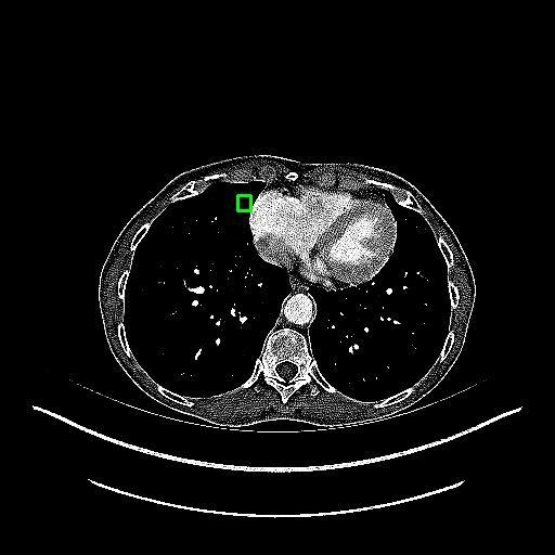
assistant:  This CT scan of the chest evaluates the lungs, heart, and upper spine, with particular attention to a region of interest in the central, upper-middle portion of the lungs.

Key observations:
- The region of interest is small in size but exhibits an unusual appearance, raising concern for a possible lesion.
- This abnormality appears to involve or affect the surrounding lung tissue.
- Its proximity to the heart and major blood vessels suggests a potential interaction between the lesion and these structures.

Further clinical correlation is required to determine the nature of this finding and its clinical significance.
|
30
|
medtrinity_conv_1__00000029
|
|
user: Summarize the content of the image as if it were an alternative text (alt text) for accessibility. <|img1|>
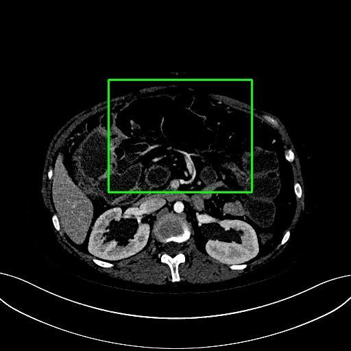
assistant:  The image is a transverse CT scan of the abdomen, displaying key abdominal organs including the liver, spleen, kidneys, and segments of the bowel.

In the central, upper-middle region of the scan, a lesion is observed within the liver. This area occupies approximately 13.1% of the total image area and appears distinct from the surrounding hepatic tissue.

The lesion may be:
- A localized hepatic abnormality, or
- Related to adjacent organs or structures, potentially indicating a systemic condition.

Its presence warrants further evaluation, as it could impact or be influenced by nearby anatomical structures.
|
31
|
medtrinity_conv_1__00000030
|
|
user: Create an image caption that would help someone understand the picture without seeing it. <|img1|>
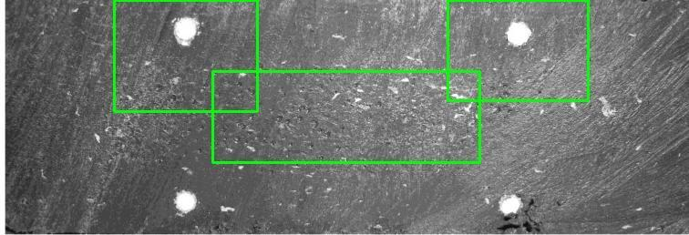
assistant:  The image is a magnetic resonance imaging (MRI) scan of the brain, displaying a cross-sectional view of cerebral structures.

- A region of interest is centered in the upper-middle portion of the image, covering approximately **8.7%** of the total area.
- This region is highlighted and shows an **abnormality** in brain tissue, evident through altered texture and intensity compared to surrounding areas.
- These changes suggest a **potential pathology**, such as a lesion, tumor, or other neurological condition.
- Due to its central location, the abnormality may be interacting with adjacent brain structures—either influencing their function or being affected by them.

Further clinical evaluation is recommended to determine the exact nature of the abnormality.
|
32
|
medtrinity_conv_1__00000031
|
|
user: Provide an objective description of what appears in the image. <|img1|>
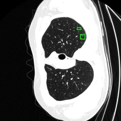
assistant:  The image is a high-resolution computed tomography (HRCT) scan of the chest, displaying the lungs, airways, and surrounding thoracic structures—without any medical devices.

A region of interest is visible in the right-center horizontally and upper-middle vertically, occupying approximately 0.1% of the total area. This region exhibits an unusual texture that may suggest signs of COVID-19 infection, including:

- Ground-glass opacity
- Consolidation
- Other atypical pulmonary features

The location of this abnormality is adjacent to the lung parenchyma, indicating a possible connection with surrounding lung tissue. This could reflect a localized area of infection or inflammation, potentially part of the broader disease process affecting the lungs.
|