| # | key | img1.png | txt |
|---|
1
|
ct2__00000000
|
|
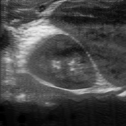
user: Can you specify the location of the kidney in this image: <|img1|>?
assistant:  The kidney's location is identified as mostly center.
|
2
|
ct2__00000001
|
|
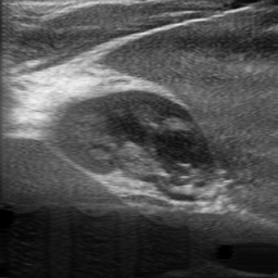
user: Identify the kidney's location in this image: <|img1|>.
assistant:  Scans show the kidney located mostly center.
|
3
|
ct2__00000002
|
|
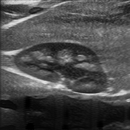
user: Show me where the kidney is located in this image: <|img1|>.
assistant:  The position of the kidney is primarily mostly center, and center-left. This is an abdominal ultrasound using a curvilinear transducer in B-mode.

Visible Organs and Structures: The image shows a transverse section of the kidney, likely the right kidney, which is well-visualized.

Features of the Kidney:
- Size: The kidney appears within normal limits; however, exact measurements can't be determined from the image alone.
- Shape: The organ maintains a regular bean-shaped contour without visible deformities.
- Texture (echotexture): The renal cortex is hypoechoic relative to the liver, with a distinguishable corticomedullary differentiation. The renal sinus appears hyperechoic.
- Borders: The kidney has smooth and well-defined borders.
- Symmetry: Symmetry can't be assessed with a single kidney in view.

Additional Findings:
- Fluid Presence: No abnormal fluid collections are observed around the kidney.
- Artifacts: Acoustic shadowing is noted posterior to the renal medulla, which is typical and expected.
- Masses/Lesions: No masses or lesions are visible.
- Vascular Features: Not applicable in this B-mode image as Doppler was not used.

Gray Scale Features: No significant hyperechoic or hypoechoic abnormalities present that could suggest pathology.

Dynamic Features: Not applicable, as this is a static image.

Image Quality and Limitations: Image quality is good, with clear visualization of the renal structures. No significant limitations such as bowel gas interference are noted.

Patient Demographic Context: Patient age and sex are not provided, which might contribute to more precise evaluation.

Impression/Conclusion: The ultrasound of the kidney shows normal morphology and echotexture. No signs of hydronephrosis, masses, or significant abnormalities are present. Further evaluation might be necessary if clinical suspicion persists.
|
4
|
ct2__00000003
|
|
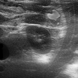
user: Where is the kidney situated in this image: <|img1|>?
assistant:  According to the scan, the kidney is located center. This is an abdominal ultrasound using a curvilinear transducer in B-mode.

Visible Organs and Structures: The image primarily shows the kidney, which is well-visualized. Adjacent tissue and possibly the liver or other abdominal structures are partially visible.

Features of Each Organ/Structure:
- Kidney:
 - Size: Appears within normal range; measurements not available.
 - Shape: Regular renal contour.
 - Texture (echotexture): The parenchyma is primarily homogeneous with some hypoechoic regions corresponding to normal renal pyramids.
 - Borders: Smooth and well-defined.
 - Symmetry: Cannot assess symmetry from a single image.

Additional Findings:
- Fluid Presence: No significant fluid collections are seen in the perinephric area.
- Artifacts: There are no prominent artifacts affecting image quality.
- Masses/Lesions: No masses or lesions identified.
- Vascular Features: No Doppler applied, so vascular flow is not assessed.

Gray Scale and Doppler Features:
- Hyperechoic regions may indicate the renal sinus and fibrofatty tissue. No Doppler findings as Doppler mode was not used.

Dynamic Features:
- Organ motion is not observable in this still image; no respiratory or valvular motion noted.

Image Quality and Limitations: Image quality is good. However, full interpretation is limited by the lack of additional planes and Doppler data.

Patient Demographic Context: No patient age or sex is available for context.

Impression/Conclusion: The kidney appears normal in size and echotexture with no evident abnormalities. For a comprehensive evaluation, additional images and possibly Doppler studies are recommended.
|
5
|
ct2__00000004
|
|
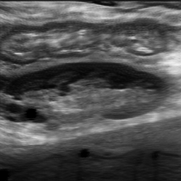
user: Could you point out the location of the kidney in this image: <|img1|>?
assistant:  The kidney's placement is described as center-right, center-left.
|
6
|
ct2__00000005
|
|
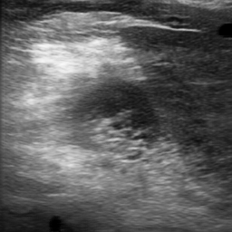
user: Highlight the location of the kidney in this image: <|img1|>.
assistant:  Ultrasound findings indicate the kidney is center.
|
7
|
ct2__00000006
|
|
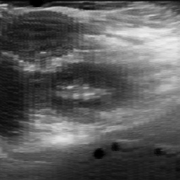
user: Can you specify the location of the kidney in this image: <|img1|>?
assistant:  The position of the kidney is primarily mostly center. Abdominal ultrasound using a curvilinear transducer in B-mode.

Visible Organs and Structures: The liver is visible and well-visualized in the image. Adjacent structures such as the gallbladder and kidneys are not clearly defined due to the focus on the liver.

Features of Each Organ/Structure:
- Liver:
 - Size: Appears normal, though specific measurements cannot be assessed from the image alone.
 - Shape: Regular shape without visible deformities.
 - Texture (echotexture): The liver demonstrates a homogeneous echotexture with consistent echo patterns across the tissue.
 - Borders: Smooth borders are observed, indicating no significant irregularity.
 - Symmetry: Not applicable as this is a single organ view.

Additional Findings:
- Fluid Presence: No significant fluid collections or ascites are noted adjacent to the liver.
- Artifacts: Minimal shadowing observed, mainly at the hepatic borders, likely due to overlying rib structures.
- Masses/Lesions: No discrete masses or lesions are observed within the liver parenchyma.
- Vascular Features: Not prominently visible in this image; Doppler mode would be required to assess vascularity effectively.

Gray Scale and Doppler Features:
Hyperechoic regions are not distinctly evident, indicating uniformity in tissue density. Hypoechoic or anechoic regions that may suggest cysts or lesions are absent.

Dynamic Features:
- Organ Motion: As this is a static image, organ motion cannot be assessed.
- Valvular Motion: Not applicable for abdominal ultrasound.

Image Quality and Limitations: The image quality appears good, with a clear depiction of the liver's structure. Minor artifact presence is noted, mainly due to anatomical interference like bowel gas or bone.

Patient Demographic Context: Information on age and sex is not available for this image.

Impression/Conclusion: The ultrasound image presents a normal-appearing liver with homogeneous echotexture and smooth borders. No pathologic findings such as masses or fluid collections are observed. Further evaluation with Doppler ultrasound could provide additional insight into vascular characteristics if clinically indicated.
|
8
|
ct2__00000007
|
|
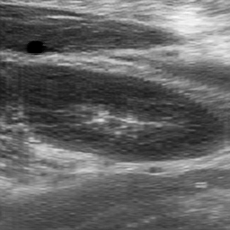
user: Which part of this image, <|img1|>, shows the kidney?
assistant:  Detected kidney positioning: center-right, center-left.
|
9
|
ct2__00000008
|
|
user: Please show the position of the kidney in this image: <|img1|>.
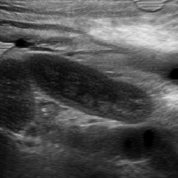
assistant:  The current scan shows the kidney positioned mostly center, and center-right.
|
10
|
ct2__00000009
|
|
user: Can you specify the location of the kidney in this image: <|img1|>?
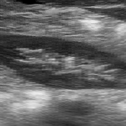
assistant:  The kidney is primarily found center-left, center-right.
|
11
|
ct2__00000010
|
|
user: Highlight the location of the kidney in this image: <|img1|>.
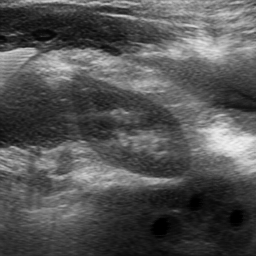
assistant:  The kidney's ultrasound indicates it is mostly center. This is an abdominal ultrasound using a curvilinear transducer in B-mode.

Visible Organs and Structures: The visible structures include the kidney and adjacent tissues. The kidney is well-visualized.

Features of Each Organ/Structure:
- Kidney:
 - Size: The kidney appears normal in size based on the visible portion.
 - Shape: The shape is regular.
 - Texture (echotexture): The renal cortex displays heterogeneous echotexture with hyperechoic regions indicating possible renal pyramids.
 - Borders: The borders are smooth and well-defined.
 - Symmetry: Unable to assess symmetry as only one kidney is visible.

Additional Findings:
- Fluid Presence: No significant fluid collections are visible in the perirenal area.
- Artifacts: There is some degree of posterior acoustic enhancement, a common feature beneath fluid-filled spaces.
- Masses/Lesions: No masses or lesions are visible.
- Vascular Features: No Doppler assessment is included, so vascular details are unavailable.

Gray Scale and Doppler Features:
- Hyperechoic regions within the kidney may indicate renal pyramids or sinus fat, which is normal. No Doppler information on flow patterns or velocities.

Dynamic Features: Organ motion is not assessed due to the static nature of the image.

Image Quality and Limitations: The image quality is good; however, the assessment is limited by the absence of Doppler data and the presence of bowel gas. Only partial visualization of the kidney is available.

Patient Demographic Context: No demographic information, such as patient age or sex, is provided.

Impression/Conclusion: This ultrasound image shows a normal-appearing kidney with regular shape and homogenous echotexture. No abnormalities, such as masses or fluid collections, are detected in this view. Further longitudinal views and Doppler evaluations would provide a more comprehensive assessment.
|
12
|
ct2__00000011
|
|
user: Please show the position of the kidney in this image: <|img1|>.
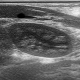
assistant:  This scan suggests the kidney is center-right, center-left.
|
13
|
ct2__00000012
|
|
user: Where can the kidney be found in this image: <|img1|>?
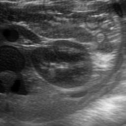
assistant:  Imaging places the kidney mostly center. This is an abdominal ultrasound using a curvilinear transducer in B-mode. The scan shows the following visible organs and structures:

1. Kidney:
 - Size: Appears normal; unable to provide exact measurements from the image.
 - Shape: Regular, with no noted deformities.
 - Texture (echotexture): Heterogeneous, typically with a distinguishable cortex and medulla.
 - Borders: Smooth and well-defined.
 - Symmetry: Evaluation of symmetry is not feasible from a single image without seeing both kidneys.

2. Fluid and Artifacts:
 - Anechoic regions likely indicating the presence of urinary structures or fluid collections.
 - Posterior enhancement suggests the fluid presence as expected in the renal collecting system.
 - Minor acoustic shadowing is visible, potentially due to renal sinus fat or calculi; however, no clear calculi seen in this image.

3. Other Structures:
 - Intestinal gas or fecal content causes acoustic shadowing, which limits visualization of other structures behind it.

4. Image Quality and Limitations:
 - The image quality is fair. Limitations include reduced visibility due to bowel gas, affecting the clarity of posterior structures.

5. Patient Demographic Context:
 - No patient age or sex provided, so unable to correlate with specific demographic characteristics.

6. Impression/Conclusion:
 - The kidney appears normal in echotexture and size, with no clear signs of hydronephrosis or masses. The anechoic areas and posterior enhancement are consistent with normal renal physiology, but further images or clinical correlation are recommended for comprehensive assessment. There are no acute abnormalities apparent in this single image.
|
14
|
ct2__00000013
|
|
user: Locate the kidney in this image: <|img1|>.
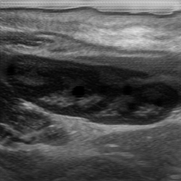
assistant:  Detected kidney positioning: center-right, center-left.
|
15
|
ct2__00000014
|
|
user: Identify the kidney's location in this image: <|img1|>.
assistant:  The kidney's position appears to be center-left, center-right.
|
16
|
ct2__00000015
|
|
user: Where can the kidney be found in this image: <|img1|>?
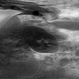
assistant:  The kidney is located center.
|
17
|
ct2__00000016
|
|
user: Can you identify the location of the kidney in this image: <|img1|>?
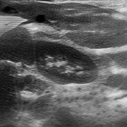
assistant:  The kidney is primarily found mostly center. This image appears to be of an abdominal ultrasound, likely using a linear transducer in B-mode.

Visible Organs and Structures: The image shows part of the kidney which is well-visualized. Additional soft tissue structures are visible, but their precise identification is unclear without annotation.

Features of Each Organ/Structure: The kidney is visible:
- Size: Cannot determine size without scale, but appears normal in relation to surrounding structures.
- Shape: Regular shape.
- Texture (echotexture): The kidney has a heterogeneous echotexture with identifiable cortical and medullary differentiation.
- Borders: Smooth and well-defined.
- Symmetry: As only one kidney is visible, symmetry cannot be assessed.

Additional Findings:
- Fluid Presence: No significant fluid collection observed.
- Artifacts: Some shadowing artifacts might be present due to dense structures.
- Masses/Lesions: No masses or lesions are clearly observed in this image.
- Vascular Features: Not applicable as no Doppler mode is used.

Gray Scale and Doppler Features: The cortex shows expected hyperechoic patterns, with the medulla appearing more hypoechoic. No Doppler features presented.

Dynamic Features: Static image provided, so no dynamic features can be assessed.

Image Quality and Limitations: The quality appears to be fair to good. One limitation is the single plane image, which may omit other features of interest such as vascular flow or anatomical anomalies.

Patient Demographic Context: No age or sex information available for the patient.

Impression/Conclusion: This is an abdominal ultrasound image showing a segment of the kidney with normal appearance in terms of echotexture and borders. No masses, significant lesions, or fluid collections are apparent. If clinical concern persists, complete both lateral and transverse scans of the kidneys, possibly with Doppler, to ensure comprehensive assessment.
|
18
|
ct2__00000017
|
|
user: Can you highlight where the kidney is in this image: <|img1|>?
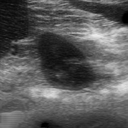
assistant:  According to the scan, the kidney is located mostly center.
|
19
|
ct2__00000018
|
|
user: Locate the kidney in this image: <|img1|>.
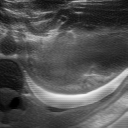
assistant:  The kidney appears to be situated center.
|
20
|
ct2__00000019
|
|
user: Could you point out the location of the kidney in this image: <|img1|>?
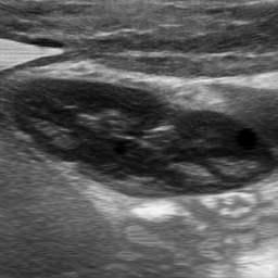
assistant:  The kidney's position appears to be center-right, center-left.
|
21
|
ct2__00000020
|
|
user: Which part of this image, <|img1|>, shows the kidney?
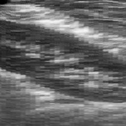
assistant:  According to the scan, the kidney is located center-left.
|
22
|
ct2__00000021
|
|
user: Where is the kidney situated in this image: <|img1|>?
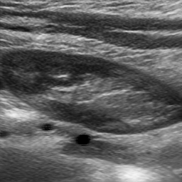
assistant:  Scans reveal the kidney to be positioned center-right, center-left. This image appears to be an ultrasound of a kidney.

Abdominal ultrasound using a curvilinear transducer, likely in B-mode.

Visible Organs and Structures: The kidney is well-visualized with identifiable surrounding renal tissue.

Features of Each Organ/Structure:
- Size: Not specified due to lack of scale, but no evident abnormal enlargement or reduction.
- Shape: Regular, maintaining typical renal contours.
- Texture (echotexture): The kidney displays a normal, heterogeneous echotexture, typical of renal tissue with cortical and medullary differentiation.
- Borders: Smooth and well-defined.
- Symmetry: Only one kidney is visible, so symmetry cannot be assessed.

Additional Findings:
- Fluid Presence: No visible perirenal fluid collections or hydronephrosis.
- Artifacts: Some acoustic shadowing is visible posterior to dense structures, typical and expected.
- Masses/Lesions: No detectable masses or lesions within the kidney.
- Vascular Features: Not assessed in this image as there is no Doppler mode apparent.

Gray Scale and Doppler Features:
- No significant hyperechoic or hypoechoic regions indicative of abnormal findings.

Dynamic Features:
- Not applicable with a static image for assessing motion.

Image Quality and Limitations: Image quality is good, but without patient details such as age and sex, complete context can't be provided.

Patient Demographic Context: Not available.

Impression/Conclusion: The kidney appears normal with no remarkable ultrasonographic abnormalities evidenced in this image. Further clinical correlation and additional imaging, as necessary, could be recommended based on the clinical context.
|
23
|
ct2__00000022
|
|
user: Highlight the location of the kidney in this image: <|img1|>.
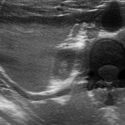
assistant:  The kidney's placement is described as center.
|
24
|
ct2__00000023
|
|
user: Could you point out the location of the kidney in this image: <|img1|>?
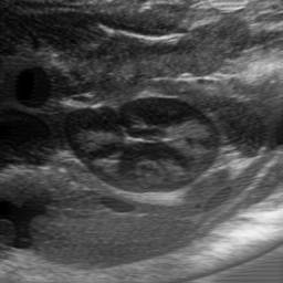
assistant:  The current scan shows the kidney positioned mostly center. This is an abdominal ultrasound using a curvilinear transducer in B-mode.

Visible Organs and Structures: The kidney is well-visualized, and part of the bowel is visible in the periphery.

Features of Each Organ/Structure:
- Kidney:
 - Size: Appears normal, but a measurement is not provided.
 - Shape: Regular shape, no deformities noted.
 - Texture (echotexture): Shows typical kidney echotexture, homogeneous.
 - Borders: Smooth and well-defined.
 - Symmetry: Only one kidney is visualized for assessment.

Additional Findings:
- Fluid Presence: No fluid collections are observed around the kidney.
- Artifacts: Mild shadowing commonly associated with bowel gas.
- Masses/Lesions: No masses or lesions detected within the kidney.
- Vascular Features: Not assessed as Doppler mode is not used in this image.

Gray Scale and Doppler Features: The renal cortex is hyperechoic compared to the medulla, as expected. No Doppler features are evaluated because the mode is not used here.

Dynamic Features: Static image, so no dynamic features such as organ motion are assessed.

Image Quality and Limitations: Image quality is good, but there is some shadowing from bowel gas, which may limit visualization of adjacent structures.

Patient Demographic Context: Not provided in the description.

Impression/Conclusion: The ultrasound image shows a normal-appearing kidney with clearly defined architecture and no abnormal findings such as masses, cysts, or fluid collections. Further evaluation with Doppler or additional imaging might be required for comprehensive assessment, particularly of vascular features.
|
25
|
ct2__00000024
|
|
user: In this image, <|img1|>, where is the kidney located?
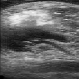
assistant:  The kidney's placement is described as center-left.
|
26
|
ct2__00000025
|
|
user: Where can the kidney be found in this image: <|img1|>?
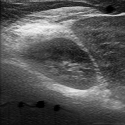
assistant:  Scans reveal the kidney to be positioned mostly center.
|
27
|
ct2__00000026
|
|
user: In this image, <|img1|>, where is the kidney located?
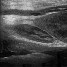
assistant:  Imaging places the kidney mostly center. This is an abdominal ultrasound using a linear transducer, taken in B-mode.

Visible Organs and Structures: The image prominently shows a transverse section of the kidney. The renal cortex, medulla, and renal pelvis are well-visualized.

Features of Each Organ/Structure:
- Kidney:
 - Size: The kidney appears to be of normal size, though specific measurements aren't provided.
 - Shape: The kidney maintains a regular shape with no apparent deformities.
 - Texture (echotexture): The renal cortex appears homogeneous and is hypoechoic compared to the surrounding structures. The medulla is relatively hyperechoic.
 - Borders: The borders of the kidney are smooth and well-defined.
 - Symmetry: Unable to assess symmetry as only one kidney is visible in this image.

Additional Findings:
- Fluid Presence: No abnormal fluid collections are noted around the kidney.
- Artifacts: There is no significant shadowing or enhancement impacting the image quality.
- Masses/Lesions: No masses or lesions are evident within this view.
- Vascular Features: Doppler assessment not available in this image, so no vascular features can be described.

Gray Scale and Doppler Features: The kidney's hypoechoic cortex and hyperechoic medulla are typical findings. Without Doppler, no flow patterns can be assessed.

Dynamic Features: No dynamic functions can be appreciated in the static image.

Image Quality and Limitations: The image quality is good with clear visualization of renal structures. Interpretation isn't hampered by positioning or adjacent bowel gas.

Patient Demographic Context: The patient's age and sex are not provided with the image.

Impression/Conclusion: The kidney appears normal in size, shape, and echotexture, with no visualized abnormalities. Consider follow-up if clinically indicated or if additional views are required for comprehensive evaluation.
|
28
|
ct2__00000027
|
|
user: Where can the kidney be found in this image: <|img1|>?
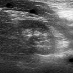
assistant:  Ultrasound findings indicate the kidney is mostly center.
|
29
|
ct2__00000028
|
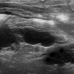
|
user: Please show the position of the kidney in this image: <|img1|>.
assistant:  The current scan shows the kidney positioned center.
|
30
|
ct2__00000029
|
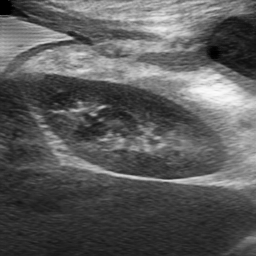
|
user: Show me where the kidney is located in this image: <|img1|>.
assistant:  Detected kidney positioning: center-right.
|
31
|
ct2__00000030
|
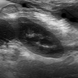
|
user: Show me where the kidney is located in this image: <|img1|>.
assistant:  Observed kidney position: mostly center. This image appears to be an abdominal ultrasound using a curvilinear transducer in B-mode.

Visible Organs and Structures: The kidney is well-visualized in the image. Adjacent structures such as the liver or spleen may be visible but are not clearly defined here.

Features of the Kidney:
- Size: The kidney appears to be of normal size, but specific measurements are not available.
- Shape: The kidney maintains a regular shape.
- Texture (echotexture): The kidney shows a heterogeneous echotexture with normal corticomedullary differentiation.
- Borders: The borders of the kidney are smooth and well-defined.
- Symmetry: A single kidney is visualized, so symmetry cannot be assessed.

Additional Findings:
- Fluid Presence: There is no visible fluid collection around the kidney.
- Artifacts: Some shadowing may be present typically due to bowel gas.
- Masses/Lesions: No obvious masses or lesions are identified.
- Vascular Features: Doppler assessment is not available in this image.

Gray Scale Features:
- The kidney parenchyma appears mildly hyperechoic relative to normal tissue, which can be clinically insignificant without additional contextual information.

Dynamic Features:
- Static image; no dynamic features such as organ motion can be assessed.

Image Quality and Limitations: Image quality seems fair, but there might be limitations due to potential overlying bowel gas or patient positioning affecting visualization.

Patient Demographic Context: Not available in the provided image.

Impression/Conclusion: The ultrasound demonstrates a normal-appearing kidney with no signs of enlargement, mass, or significant abnormality. Further evaluation with Doppler or additional imaging could offer more insights into vascular or functional concerns if clinically indicated. If no clinical symptoms are presented, findings are likely incidental.
|
32
|
ct2__00000031
|
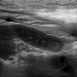
|
user: Show me where the kidney is located in this image: <|img1|>.
assistant:  Scans show the kidney located mostly center, and center-right.
|