| # | key | img1.png | txt |
|---|
1
|
covid_us__00000000
|
|
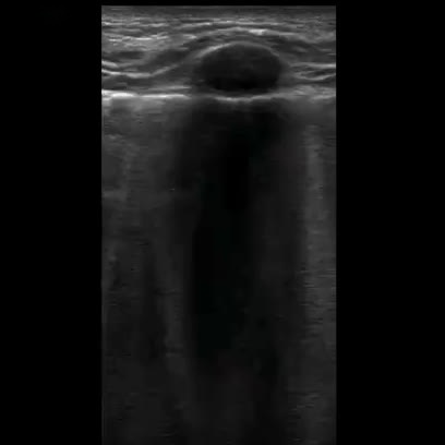
user: What can be observed in <|img1|>?
assistant:  The lung is visualized here with a linear probe. The patient shows symptoms of Pneumonia. The computed LUSS is 1. Soft tissue ultrasound using a linear transducer in B-mode.

Visible Organs and Structures: The image primarily shows subcutaneous tissue with a visible circular structure, likely a lesion.

Features of Each Organ/Structure:
- Size: The circular structure appears moderately sized, but precise measurements cannot be determined from the image alone.
- Shape: The lesion is round and appears regular in shape.
- Texture (echotexture): The lesion exhibits a hypoechoic center with some anechoic areas, suggesting fluid content.
- Borders: The borders of the lesion are smooth and well-defined.
- Symmetry: The lesion is solitary, so symmetry is not applicable.

Additional Findings:
- Fluid Presence: Anechoic areas suggest possible fluid collection within the lesion.
- Artifacts: Posterior acoustic enhancement is evident, indicating increased echogenicity beyond the lesion due to fluid presence.
- Masses/Lesions: The described hypoechoic lesion could represent a cyst or an abscess depending on clinical correlation and patient presentation.
- Vascular Features: Not assessed due to lack of Doppler use in the image.

Gray Scale and Doppler Features: Hypoechoic and anechoic regions support the presence of fluid. No Doppler findings are available in this static image.

Dynamic Features: Not applicable in the static image provided.

Image Quality and Limitations: The image quality is good, with clear visualization of the lesion. Lack of Doppler mode limits vascular assessment.

Patient Demographic Context: Not available from the image alone.

Impression/Conclusion: The ultrasound image shows a well-defined, hypoechoic lesion with anechoic components, likely representing a cystic structure with possible fluid content. Differential considerations include a sebaceous cyst or abscess. Further clinical correlation or additional imaging may be necessary for definitive diagnosis.
|
2
|
covid_us__00000001
|
|
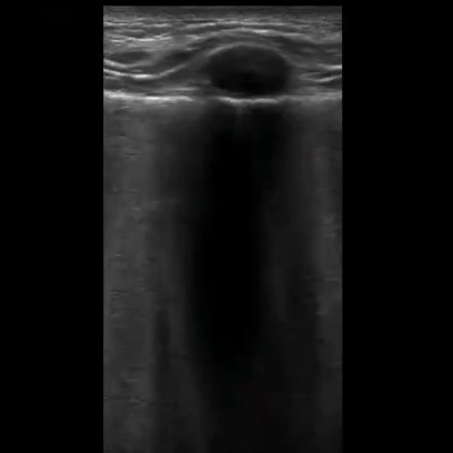
user: Describe the following image: <|img1|>
assistant:  An ultrasound image depicting lung using a linear probe. The patient is suffering from Pneumonia. A LUSS of 1 is reported. This is an ultrasound image, likely from a thyroid or superficial neck study, utilizing a linear transducer in B-mode.

Visible Organs and Structures:
The image prominently features a superficial mass, with the overlying skin and subcutaneous tissue visible. Deeper structures are not well defined.

Features of the Mass:
- Size: The mass appears well-defined but precise measurements are not possible from the image alone.
- Shape: The mass is oval or round in shape.
- Texture (echotexture): It exhibits a predominantly hypoechoic texture.
- Borders: The borders appear smooth and well-defined.
- Symmetry: No bilateral structures are available for comparison.

Additional Findings:
- Fluid Presence: There is no evidence of fluid collection or cystic components around the mass.
- Artifacts: Posterior acoustic enhancement is noted, which is typical for hypoechoic structures.
- Masses/Lesions: The lesion displays homogeneous echotexture, suggesting a benign nature, although further clinical correlation and possibly Doppler analysis would help.
- Vascular Features: Doppler was not applied, so no vascular information is provided.

Gray Scale and Doppler Features:
- Hypoechoic regions typically indicate fluid collections, solid organs, or masses. Clinical correlation is necessary to determine significance.
- No Doppler features are visible, hence no information on flow patterns or velocities is available.

Dynamic Features:
- Not applicable as the image is a static B-mode ultrasound without dynamic assessment.

Image Quality and Limitations:
- The image quality is good, with clear delineation of the mass. There are no noted limitations such as bowel gas or patient movement.

Patient Demographic Context:
- No specific patient age or sex details are provided.

Impression/Conclusion:
The ultrasound image reveals a single, well-defined hypoechoic mass, likely benign. The presence of posterior acoustic enhancement suggests a solid consistent lesion. Differential diagnoses could include a benign thyroid nodule or lipoma, but further evaluation with Doppler ultrasound and clinical correlation is recommended.
|
3
|
covid_us__00000002
|
|
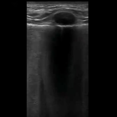
user: What does <|img1|> show?
assistant:  An ultrasound image depicting lung using a linear probe. The patient is suffering from Pneumonia. The computed LUSS is 1. This is an ultrasound image that appears to be taken with a linear transducer, typically used for superficial structures. The mode is likely B-mode.

Visible Organs and Structures: The image prominently shows subcutaneous tissue and a round structure that may be a cyst or nodule.

Features of Each Organ/Structure:
- Round Structure:
 - Size: Appears to be small and round; exact measurements are not provided.
 - Shape: Regular, round shape.
 - Texture: Anechoic center, suggesting a fluid-filled structure like a cyst.
 - Borders: Smooth, well-defined borders.
 - Symmetry: Not applicable as it is a single structure.

Additional Findings:
- Fluid Presence: The anechoic center indicates the presence of fluid.
- Artifacts: There is posterior acoustic enhancement, suggesting a cystic nature.
- Masses/Lesions: The structure could be a cyst; other possibilities depend on clinical context.
- Vascular Features: Not applicable in this image as Doppler is not used.

Gray Scale and Doppler Features: The hyperechoic borders and anechoic center suggest a fluid-filled structure with posterior enhancement. Without Doppler, no vascular features can be assessed.

Dynamic Features: Not applicable as this is a static image.

Image Quality and Limitations: The image quality appears good, with clear delineation of the structure and no evident obscuring factors such as bowel gas.

Patient Demographic Context: Not provided in the image.

Impression/Conclusion: The image shows a well-defined anechoic structure with posterior enhancement, consistent with a simple cyst. Further correlation with patient history and possibly additional imaging is recommended for a definitive diagnosis.
|
4
|
covid_us__00000003
|
|
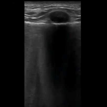
user: What is depicted in the image <|img1|>?
assistant:  This ultrasound highlights lung with the help of a linear probe. The patient is diagnosed with Pneumonia. The calculated LUSS value is 1. This is a thyroid ultrasound using a linear transducer in B-mode.

Visible Organs and Structures: The thyroid gland is visible, specifically showing part of the parenchyma. The focus is on a nodule within the thyroid tissue.

Features of Each Organ/Structure:
- Nodule (Thyroid):
 - Size: Appears localized, precise measurements are not provided.
 - Shape: Oval and regular.
 - Texture (echotexture): Hypoechoic compared to the surrounding thyroid tissue.
 - Borders: Smooth and well-defined.
 - Symmetry: Only one side is visible, so symmetry cannot be assessed.

Additional Findings:
- Fluid Presence: Anechoic area suggests the presence of fluid, possibly indicating a cystic component.
- Artifacts: There is posterior acoustic enhancement beneath the nodule, a common artifact with cystic structures.
- Masses/Lesions: The nodule is likely cystic due to its anechoic center and posterior enhancement.

Gray Scale and Doppler Features:
- Hypoechoic regions suggest possible cystic nature; no Doppler features are visible in this image.

Dynamic Features:
- No dynamic features, such as motion artifacts, are observed.

Image Quality and Limitations: Image quality is good, but only part of the thyroid is visible. The presence of a single nodule limits assessment of the entire gland. Adequate positioning, minimal shadowing from surrounding structures.

Patient Demographic Context: Patient age and sex are not available, which could impact differential diagnosis considerations.

Impression/Conclusion: The ultrasound shows a well-defined, hypoechoic, likely cystic nodule in the thyroid gland, possibly benign. Further correlation with patient history, lab tests (e.g., thyroid function tests), and additional imaging or follow-up may be warranted to rule out malignancy.
|
5
|
covid_us__00000004
|
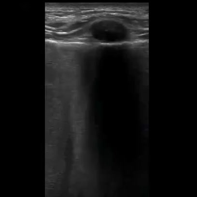
|
user: Please summarize the details of <|img1|>.
assistant:  A linear probe captured this ultrasound of lung. Abnormalities suggestive of Pneumonia are seen. The calculated LUSS value is 1. This is a soft tissue ultrasound, likely using a linear transducer in B-mode.

Visible Organs and Structures: The image primarily shows subcutaneous tissue with an anechoic region suggestive of a cyst or fluid collection. The skin and superficial tissue layers are well-visualized.

Features of Each Organ/Structure:
- Anechoic Region (possible cyst):
 - Size: Appears regular; exact measurements are not provided.
 - Shape: Round and well-defined.
 - Texture (echotexture): Anechoic, indicating a fluid-filled structure.
 - Borders: Smooth and well-defined.
 - Symmetry: Not applicable as this is a single structure.

Additional Findings:
- Fluid Presence: There is a clear anechoic area suggesting a fluid collection, possibly a cyst.
- Artifacts: Posterior acoustic enhancement is seen beneath the anechoic area, which is typical for fluid collections, indicating increased echo transmission.
- Masses/Lesions: The anechoic region itself may represent a mass, likely a benign cyst given its characteristics.
- Vascular Features: No vascular flow is noted in this area.

Gray Scale and Doppler Features:
- Hyperechoic and hypoechoic regions are not prominent apart from the mentioned structures.
- No Doppler flow noted, suggesting the structure is not vascular.

Dynamic Features: No dynamic features or motion applicable in this image.

Image Quality and Limitations: Image quality appears good with clear visualization of the anechoic area and surrounding tissues. No significant limitations visible but note that detailed characterization may require correlation with clinical findings and possible further imaging.

Patient Demographic Context: No specific patient demographic information is available from this image.

Impression/Conclusion: The ultrasound image reveals an anechoic fluid-filled structure in the soft tissues, likely consistent with a simple cyst. There is posterior acoustic enhancement without signs of internal complexity or vascularity. Clinical correlation is recommended to rule out other potential causes of fluid collection, although features are most suggestive of a benign cyst.
|
6
|
covid_us__00000005
|
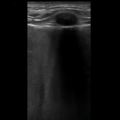
|
user: <|img1|> Describe this image
assistant:  The lung is visualized here with a linear probe. The results are consistent with a diagnosis of Pneumonia. This image reflects a LUSS of 1. This is most likely a soft tissue ultrasound using a linear transducer in B-mode.

Visible Organs and Structures: A superficial mass in the subcutaneous tissue is visible, with surrounding soft tissue structures. The depth and tissue penetration suggest a superficial location.

Features of Each Organ/Structure:
- Size: The mass appears round and small. Exact measurements are not provided, but it seems to occupy a localized area in the superficial tissue.
- Shape: Regular, with a round appearance.
- Texture (echotexture): The mass is mostly anechoic internally, indicating fluid content.
- Borders: The borders are smooth and well-defined.
- Symmetry: Not applicable as only a single focal mass is visible.

Additional Findings:
- Fluid Presence: The mass appears anechoic, consistent with a cystic structure, indicating a potential fluid collection.
- Artifacts: Acoustic shadowing is evident, likely due to the solid nature or calcifications within the mass.
- Masses/Lesions: A single round anechoic mass is noted; size and precise location are unspecified.
- Vascular Features: Doppler assessment is not visible, so vascularity cannot be assessed.

Gray Scale and Doppler Features:
- Hyperechoic, hypoechoic, and anechoic regions are consistent with a simple cyst due to the mass's anechoic nature.
- Doppler findings are not available.

Dynamic Features:
- Organ motion or other dynamic features are not assessable due to the static nature of the image provided.

Image Quality and Limitations: The image quality appears good with clear demarcation of the mass. However, the static image limits the assessment of vascularity and other dynamic features.

Patient Demographic Context: Age and sex of the patient are not specified, which may influence the differential diagnosis.

Impression/Conclusion: The ultrasound reveals a well-defined, round, anechoic mass in the subcutaneous tissue with posterior acoustic shadowing, likely a sebaceous cyst or lipoma. Further clinical correlation and dynamic assessment with Doppler imaging are recommended for a comprehensive evaluation.
|
7
|
covid_us__00000006
|
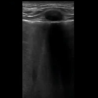
|
user: What is depicted in the image <|img1|>?
assistant:  Lung imaged with a linear probe. This shows clear signs of Pneumonia. A LUSS of 1 is reported. This is a soft tissue or musculoskeletal ultrasound, likely using a linear transducer in B-mode.

Visible Organs and Structures: The image prominently displays subcutaneous tissue and potentially part of a muscle layer. These structures appear well-visualized.

Features of Each Organ/Structure:
- Size: The subcutaneous tissue appears within normal thickness as visualized.
- Shape: The visible structures appear regular without evident deformities.
- Texture (echotexture): The subcutaneous layer is moderately echogenic, while deeper tissue layers appear hypoechoic.
- Borders: Smooth and well-defined.
- Symmetry: Not applicable to this single view.

Additional Findings:
- Fluid Presence: A visible anechoic area suggests a fluid collection, possibly a cyst or other fluid-filled structure.
- Artifacts: Posterior acoustic enhancement is noted beneath the anechoic area.
- Masses/Lesions: The anechoic area is consistent with a cystic structure, with smooth, well-defined borders.
- Vascular Features: Not applicable in this image without Doppler mode.

Gray Scale and Doppler Features: The anechoic region indicates a potential cyst or fluid collection; no Doppler assessment is available in this mode.

Dynamic Features: Not applicable as this is a static image.

Image Quality and Limitations: The image quality is good. No significant limitations are apparent, although deeper structures may be obscured by the primary area of interest.

Patient Demographic Context: Patient age and sex are not provided.

Impression/Conclusion: The ultrasound image shows a well-defined anechoic area within the soft tissue, suggestive of a cyst or fluid collection. Further clinical correlation and possibly complementary imaging could be considered for a definitive diagnosis. Large vessels and critical structures appear unaffected.
|
8
|
covid_us__00000007
|
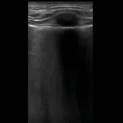
|
user: What is depicted in the image <|img1|>?
assistant:  This is an image of lung obtained using a linear probe. The patient is diagnosed with Pneumonia. A LUSS of 1 is reported. This is a soft tissue ultrasound, likely using a linear transducer in B-mode.

Visible Organs and Structures: The image appears to show superficial soft tissue structures, potentially a section of skin or subcutaneous tissue. A cystic structure is evident centrally.

Features of Each Organ/Structure:
- Cystic Structure:
 - Size: Measurement not provided, but the area is noticeable in the image.
 - Shape: Appears round and well-defined.
 - Texture (echotexture): Anechoic, indicating fluid content.
 - Borders: Smooth and well-circumscribed.
 - Symmetry: Not applicable as this is a single structure.

Additional Findings:
- Fluid Presence: A central anechoic area suggests a fluid-filled cystic structure.
- Artifacts: Acoustic enhancement is observed posterior to the cystic structure, typical of fluid-filled regions.

Gray Scale and Doppler Features:
- Hypoechoic and anechoic areas indicate potential cyst. No Doppler information provided to assess vascular flow.

Dynamic Features: The image does not provide information on dynamic features like organ motion.

Image Quality and Limitations: Image quality appears adequate for identifying the cystic structure. No significant limitations noted, but further context or views could aid comprehensive assessment.

Patient Demographic Context: Age and sex of the patient are not provided.

Impression/Conclusion: The ultrasound suggests the presence of a simple cystic structure in soft tissue, likely benign and fluid-filled, such as a sebaceous cyst. Clinical correlation and further examination are advised for definitive diagnosis.
|
9
|
covid_us__00000008
|
|
user: Give a description of <|img1|>.
assistant:  The lung is visualized here with a linear probe. The patient shows symptoms of Pneumonia. The Lung Ultrasound Score (LUSS) is 1. The image appears to be an ultrasound of a superficial structure, possibly a thyroid or soft tissue structure, using a linear transducer in B-mode.

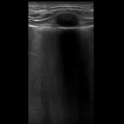
Visible Organs and Structures: A circular, well-defined structure is visible near the surface, likely representing a cystic form.

Features of Each Organ/Structure:
- Size: The structure is of moderate size, exact measurements are not visible.
- Shape: The shape is round and regular.
- Texture (echotexture): The interior is anechoic, suggesting a fluid-filled cyst.
- Borders: Borders appear smooth and well-defined.
- Symmetry: No bilateral structures present for comparison.

Additional Findings:
- Fluid Presence: The anechoic nature suggests a fluid-filled space typical of a cyst.
- Artifacts: Posterior acoustic enhancement is visible below the anechoic area, which is consistent with a fluid-filled structure.
- Masses/Lesions: The lesion is likely a simple cyst due to its characteristics, with no solid components observed.

Gray Scale and Doppler Features: The structure's anechoic nature is typical of simple cysts and clinically significant for identifying fluid-filled spaces. No Doppler flow is apparent, which aligns with the cystic nature.

Dynamic Features: Not applicable as the image is static, and no dynamic features are visible.

Image Quality and Limitations: The image quality is good for evaluating superficial structures, but limitations include the lack of measurements and patient demographic context.

Impression/Conclusion: The ultrasound image is suggestive of a superficial simple cyst, likely benign. Further clinical correlation and follow-up imaging may be considered to confirm the diagnosis. No suspicious features are noted.
|
10
|
covid_us__00000009
|
|
user: Here is an image: <|img1|>. Give a description of it
assistant:  Lung imaged with a linear probe. Abnormalities suggestive of Pneumonia are seen. LUSS is measured at 1. This is an ultrasound using a linear transducer in B-mode, likely focusing on soft tissue or superficial structures.

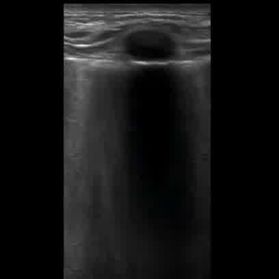
Visible Organs and Structures: The image prominently displays subcutaneous tissue and potentially underlying muscle or soft tissue. The structures beyond the initial layers are poorly visualized, likely due to artifact.

Features of Each Organ/Structure:
- Size: The visible subcutaneous layer appears within normal limits.
- Shape: Regular, with no noted deformities or asymmetry.
- Texture (echotexture): The subcutaneous tissue has a homogeneous echotexture.
- Borders: The borders are smooth and well-defined at the skin surface.
- Symmetry: Not applicable as the image is not bilateral.

Additional Findings:
- Fluid Presence: None observed in the well-visualized areas.
- Artifacts: There is a prominent shadowing artifact extending from a superficial structure, possibly indicating a foreign body or calcification.
- Masses/Lesions: None observed based on the visible portion.
- Vascular Features: Not applicable as Doppler is not used here.

Gray Scale and Doppler Features:
- Hyperechoic region at the top surface suggests a dense structure causing shadowing, such as calcification or foreign material. Anechoic shadowing extends downwards.

Dynamic Features: Not applicable as this is a still image without dynamic assessment.

Image Quality and Limitations: Image quality appears fair, hindered primarily by shadowing artifact which limits deeper visualization. This limits assessment of underlying structures.

Patient Demographic Context: Not provided.

Impression/Conclusion: The ultrasound shows a linear shadowing artifact, suggesting the presence of a dense superficial structure like a foreign body or calcification. Further evaluation with different imaging modalities may be warranted for a comprehensive assessment.
|
11
|
covid_us__00000010
|
|
user: Describe the following image: <|img1|>
assistant:  An ultrasound image depicting lung using a linear probe. The patient is suffering from Pneumonia. LUSS scoring indicates a value of 1. Soft tissue ultrasound using a linear transducer in B-mode.

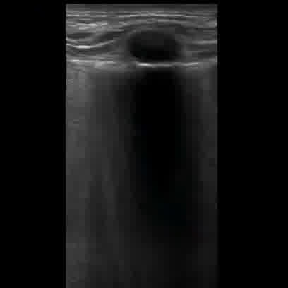
Visible Organs and Structures: The image shows subcutaneous tissue and potentially part of a small cystic structure near the surface.

Features of Each Organ/Structure:
- Subcutaneous Tissue:
 - Size: Normal.
 - Shape: Regular.
 - Texture: Homogeneous.
 - Borders: Well defined.

- Cystic Structure:
 - Size: Appears small; exact measurements not provided.
 - Shape: Round.
 - Texture: Anechoic, indicating fluid content.
 - Borders: Smooth and clearly defined.

Additional Findings:
- Fluid Presence: Anechoic region likely represents a fluid collection, consistent with a simple cyst.
- Artifacts: There is posterior acoustic enhancement beneath the cystic area, typical for fluid.
- Masses/Lesions: The cystic area is small with benign features and smooth borders.

Gray Scale and Doppler Features:
- Hypoechoic regions represent soft tissue; the anechoic area suggests a simple cyst.
- No Doppler flow is visible within the cystic area, aligning with its avascular nature.

Dynamic Features: Not applicable in the static image provided.

Image Quality and Limitations: Image quality is good with clear delineation of structures. No significant artifacts affect interpretability. However, the image doesn't display deeper tissues, possibly due to surface positioning.

Patient Demographic Context: Patient age and sex are not available.

Impression/Conclusion: The ultrasound image reveals a small, well-defined anechoic structure in the subcutaneous tissue suggestive of a simple cyst, likely benign. Further clinical correlation and, if necessary, follow-up imaging could provide additional information.
|
12
|
covid_us__00000011
|
|
user: <|img1|> Describe this image
assistant:  A linear probe captured this ultrasound of lung. The patient is diagnosed with Pneumonia. LUSS scoring indicates a value of 1. Soft tissue ultrasound using a linear transducer in B-mode.

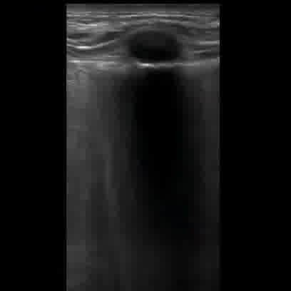
Visible Organs and Structures: The ultrasound image shows superficial skin and subcutaneous tissue. A circular anechoic structure is visible, likely a cyst or fluid collection, although deeper structures are not visible.

Features of Each Organ/Structure:
- Size: The anechoic area is approximately circular but exact measurements are not provided.
- Shape: Regular, round shape.
- Texture (echotexture): The area is anechoic, suggesting fluid content.
- Borders: Smooth, well-defined borders.
- Symmetry: Not applicable as only a single structure is visible.

Additional Findings:
- Fluid Presence: There is a prominent anechoic region suggestive of a cyst or similar fluid collection.
- Artifacts: Acoustic enhancement is present beneath the anechoic area.
- Masses/Lesions: A potential cyst is noted with the characteristics described above.

Gray Scale and Doppler Features:
- Anechoic region suggests a fluid-filled structure such as a cyst.
- Doppler features are not applicable as Doppler mode was not used.

Dynamic Features:
- Not applicable as this is a static image.

Image Quality and Limitations: Image quality is good for superficial structures, but there is limited penetration, obscuring deeper anatomy. The linear probe is optimal for superficial imaging.

Patient Demographic Context: Not provided.

Impression/Conclusion: The ultrasound image reveals a well-defined, anechoic, likely cystic structure within the superficial tissue, possibly representing a sebaceous cyst or simple fluid collection. Further clinical correlation and history are recommended for diagnosis.
|
13
|
covid_us__00000012
|
|
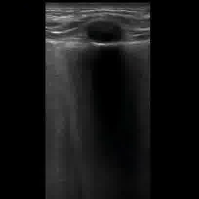
user: Describe what you see in <|img1|>.
assistant:  This ultrasound image shows lung using a linear probe. The patient shows symptoms of Pneumonia. LUSS scoring indicates a value of 1. This is a soft tissue ultrasound, likely using a linear transducer in B-mode.

Visible Organs and Structures: The image shows a section of subcutaneous tissue. A notable structure is visible in the center.

Features of Each Organ/Structure:
- Size: The central structure appears well-circumscribed; specific measurements are unavailable.
- Shape: The structure is round or oval.
- Texture (echotexture): The structure is anechoic, suggesting fluid content.
- Borders: Borders are smooth and well-defined.
- Symmetry: Not applicable as a single structure is evaluated.

Additional Findings:
- Fluid Presence: The anechoic structure suggests a simple cyst or fluid collection.
- Artifacts: Posterior acoustic enhancement is visible, which is consistent with fluid content.
- Masses/Lesions: The observed lesion appears consistent with a simple cyst.
- Vascular Features: No Doppler involved in this image, so vascular features are unavailable.

Gray Scale and Doppler Features: The anechoic region indicates fluid, likely a benign finding such as a cyst.

Dynamic Features: Motion not assessed in this static image.

Image Quality and Limitations: Image quality is good, but depth is limited to superficial tissues. There are no factors, such as bowel gas, affecting this evaluation.

Patient Demographic Context: Not provided.

Impression/Conclusion: The findings suggest a simple cystic structure in the subcutaneous tissues. Typical for benign cysts or seromas, but clinical correlation is recommended for definitive diagnosis. Further evaluation with clinical settings might be required.
|
14
|
covid_us__00000013
|
|
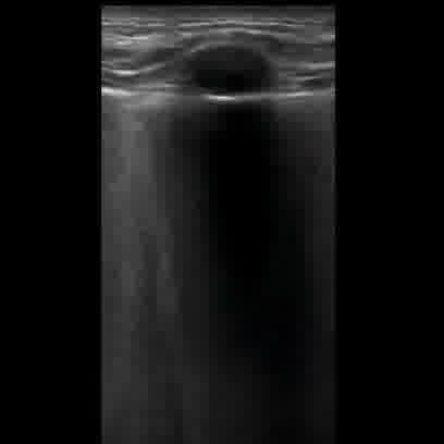
user: Here is an image: <|img1|>. Give a description of it
assistant:  Ultrasound imaging of lung performed with a linear probe. Abnormalities suggestive of Pneumonia are seen. The calculated LUSS value is 1. This is an ultrasound image using a linear transducer in B-mode, typically used for evaluating superficial structures like the thyroid, breast, or vascular access sites.

Visible Organs and Structures: The image shows superficial tissue layers, likely skin and subcutaneous tissue. A well-defined circular structure is visible near the surface.

Features of Each Organ/Structure:
- The circular structure:
 - Size: Appears as a well-defined round object; precise measurements are not provided.
 - Shape: Circular and regular.
 - Texture (echotexture): Anechoic, indicating fluid content.
 - Borders: Smooth and well-defined.

Additional Findings:
- Fluid Presence: The anechoic nature of the structure suggests it is a fluid-filled cyst.
- Artifacts: Notable posterior acoustic enhancement deep to the anechoic structure, typical of cystic formations.
- Masses/Lesions: The structure resembles a simple cyst upon initial assessment.

Gray Scale and Doppler Features: The cyst's anechoic appearance indicates a fluid collection without internal echoes, typical for simple cysts. No Doppler features observed, suggesting absence of flow within the anechoic area.

Dynamic Features: Not applicable, as this is a static image.

Image Quality and Limitations: The image quality is good, with clear visualization of superficial structures. No obscuring factors such as bowel gas are present.

Patient Demographic Context: No specific demographic details provided.

Impression/Conclusion: The image likely depicts a simple superficial cyst, given its anechoic nature and posterior enhancement. This finding is common in sites like the breast or subcutaneous regions. Further clinical correlation and possible follow-up imaging may be necessary to confirm diagnosis and monitor changes over time.
|
15
|
covid_us__00000014
|
|
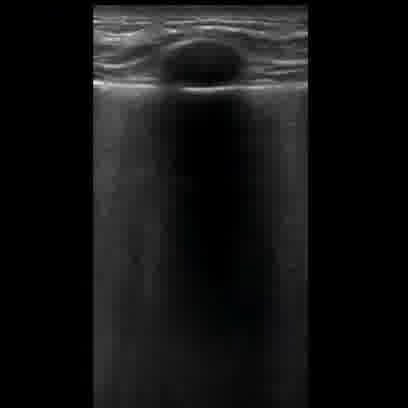
user: What does <|img1|> show?
assistant:  The lung is visualized here with a linear probe. The patient is diagnosed with Pneumonia. A LUSS of 1 is reported. This is an ultrasound image possibly performed with a linear transducer in B-mode, commonly used for examining superficial structures.

Visible Organs and Structures: The image primarily shows soft tissue with a well-defined structure near the surface, likely representing a cyst or other fluid-filled structure.

Features of Each Organ/Structure:
- Size: The structure appears as a small, rounded lesion; specific measurements are not available.
- Shape: It is circular and regular.
- Texture (echotexture): The lesion is anechoic, indicating a fluid-filled nature.
- Borders: The borders are smooth and well-defined.
- Symmetry: Not applicable, as only one structure is noted.

Additional Findings:
- Fluid Presence: The anechoic nature suggests the presence of fluid, consistent with a cyst.
- Artifacts: There is posterior acoustic enhancement, a common artifact associated with cystic structures.
- Masses/Lesions: The described lesion is consistent with a simple cyst given its anechoic nature and posterior enhancement.
- Vascular Features: No Doppler flow is visible, supporting the avascular nature of the cyst.

Gray Scale and Doppler Features:
- The anechoic appearance is clinically significant for fluid, typically found in cystic lesions.
- Doppler findings are not applicable as the image does not include Doppler mode.

Dynamic Features: Not applicable, as this is a static image and does not show any motion.

Image Quality and Limitations: The image quality is good, allowing for accurate identification of the described structure. No major limitations are noted, aside from the inability to measure the lesion directly from the image provided.

Patient Demographic Context: Not provided.

Impression/Conclusion: The ultrasound image reveals a well-defined anechoic structure with posterior acoustic enhancement, consistent with a simple cyst. No concerning features such as septations or nodularity are visible. Further clinical correlation is recommended to support this interpretation.
|
16
|
covid_us__00000015
|
|
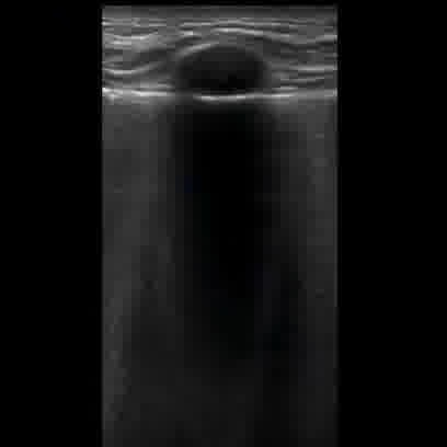
user: How would you summarize <|img1|> visually?
assistant:  This is an image of lung obtained using a linear probe. The findings indicate the presence of Pneumonia. The LUSS score is 1. This is a soft tissue ultrasound using a linear transducer in B-mode imaging.

Visible Organs and Structures: The image displays the cutaneous and subcutaneous tissue layers. Visible structures include skin and subcutaneous tissues.

Features of Each Organ/Structure:
- Size: There is a prominent round lesion present.
- Shape: The lesion is round and well-defined.
- Texture (echotexture): The lesion appears anechoic, suggesting fluid content.
- Borders: Smooth borders surrounding the lesion.
- Symmetry: Only the lesion is assessed, so symmetry is not applicable.

Additional Findings:
- Fluid Presence: The lesion is consistent with a fluid-filled collection.
- Artifacts: Posterior acoustic enhancement is present, typical for fluid-filled structures.
- Masses/Lesions: The lesion appears cystic, likely benign given its anechoic nature and smooth borders.
- Vascular Features: Not assessed in this image.

Gray Scale and Doppler Features: The anechoic lesion suggests a simple cyst, with no internal echoes indicative of complex fluid or solid components.

Dynamic Features: Not applicable in this static image.

Image Quality and Limitations: The image quality is good. No external factors such as positioning or gas obscuring interpretation.

Patient Demographic Context: Not available from the image.

Impression/Conclusion: The ultrasound image demonstrates a well-defined anechoic lesion within the subcutaneous tissue, likely representing a simple cyst. Further evaluation with Doppler and clinical correlation is recommended to confirm the diagnosis.
|
17
|
covid_us__00000016
|
|
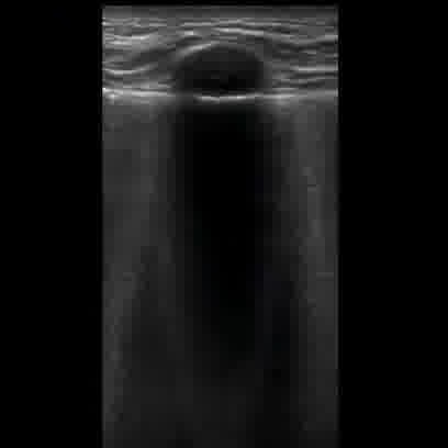
user: Here is an image: <|img1|>. Give a description of it
assistant:  An ultrasound image depicting lung using a linear probe. Abnormalities suggestive of Pneumonia are seen. A LUSS of 1 is reported. This is a soft tissue ultrasound using a linear transducer in B-mode.

Visible Organs and Structures: The image primarily shows skin, subcutaneous tissue, and a well-defined dark area, likely a cyst or fluid collection.

Features of Each Organ/Structure:
- Skin and Subcutaneous Tissue:
 - Size: Normal thickness.
 - Shape: Regular contour.
 - Texture (echotexture): Hyperechoic and homogeneous.
 - Borders: Smooth and well-defined.
 - Symmetry: Not applicable in this view.

- Cyst or Fluid Collection:
 - Size: Approximate dimensions should be measured in two planes.
 - Shape: Regular and round.
 - Texture (echotexture): Anechoic, suggesting fluid content.
 - Borders: Smooth and well-defined.

Additional Findings:
- Fluid Presence: Single anechoic area suggesting fluid collection, likely a benign cyst.
- Artifacts: Posterior acoustic enhancement is present due to the fluid-filled nature of the structure.
- Masses/Lesions: No solid masses or lesions visible.
- Vascular Features: Not assessed due to lack of Doppler imaging.

Gray Scale and Doppler Features: The anechoic region is consistent with simple fluid; no Doppler feature analysis possible here.

Dynamic Features: Not applicable; no dynamic structures visible in this static image.

Image Quality and Limitations: Image quality appears good with clear visualization of superficial structures; deeper structures are not assessed due to depth limitations and lack of deeper penetration by a linear transducer.

Patient Demographic Context: Not available, which may limit context-specific interpretation.

Impression/Conclusion: The ultrasound image suggests a regular, well-defined anechoic area consistent with a simple fluid-filled cyst, likely benign. Further clinical correlation is recommended to rule out any other potential pathology.
|
18
|
covid_us__00000017
|
|
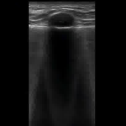
user: Here is an image: <|img1|>. Describe it
assistant:  This is an image of lung obtained using a linear probe. This shows clear signs of Pneumonia. The calculated LUSS value is 1. This is a linear transducer ultrasound, likely in B-mode, typically used for musculoskeletal or superficial soft tissue imaging.

Visible Organs and Structures: The image appears to show skin and subcutaneous tissue layers. A prominent, vertically oriented anechoic structure is visible, suggesting the presence of fluid or a cyst.

Features of Each Organ/Structure:
- Size: The anechoic structure is elongated vertically.
- Shape: Appears regular and tube-like without any noticeable deformities.
- Texture (echotexture): The structure is anechoic, consistent with fluid-filled content.
- Borders: The borders are smooth and well-defined.
- Symmetry: Not applicable as only one structure is assessed.

Additional Findings:
- Fluid Presence: The anechoic area suggests a fluid collection, possibly a cyst or a duct.
- Artifacts: The image shows posterior acoustic enhancement, a common artifact when imaging anechoic structures like cysts.
- Masses/Lesions: No solid masses are noted within the anechoic structure.
- Vascular Features: Doppler assessment would be needed to evaluate any potential flow, not visible here.

Gray Scale and Doppler Features: The anechoic region's acoustic enhancement supports its fluid nature. No Doppler flow information is provided.

Dynamic Features: Not applicable; image is static.

Image Quality and Limitations: Image quality appears good for superficial tissues, but deeper structures are not visible due to shadowing.

Patient Demographic Context: No patient age or sex provided.

Impression/Conclusion: The ultrasound image displays a well-defined, anechoic structure consistent with a benign fluid collection such as a simple cyst. Further evaluation with clinical correlation and possibly Doppler ultrasound may be needed for a comprehensive assessment.
|
19
|
covid_us__00000018
|
|
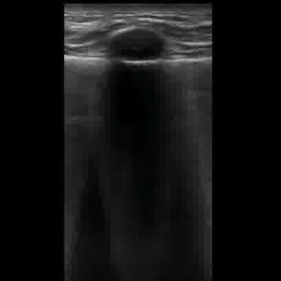
user: Describe the following image: <|img1|>
assistant:  This is an image of lung obtained using a linear probe. The findings indicate the presence of Pneumonia. The calculated LUSS value is 1. This is an ultrasound image using a linear transducer, typically utilized for superficial structures. The mode appears to be B-mode, which is used for structural assessment.

Visible Organs and Structures: The image distinctly shows superficial tissues, likely skin and subcutaneous layers. There is a noticeable dark, anechoic area suggestive of fluid or an artifact.

Features of Each Organ/Structure:
- Superficial Tissue: This area appears well-visualized, with normal looking layers above the anechoic region.
 - Size: Normal thickness for soft tissue is observed, though precise measurements are not provided.
 - Shape: The layers appear regular.
 - Texture: The tissue is homogeneous, typical for soft tissue.
 - Borders: The borders appear smooth.

Additional Findings:
- Fluid Presence: The large anechoic area could indicate a fluid collection; its shape suggests a possible cystic structure or artifact like posterior enhancement.
- Artifacts: Posterior acoustic shadowing or enhancement may be present due to the anechoic region.

Gray Scale and Doppler Features:
- Anechoic Region: The anechoic area likely indicates fluid presence and may clinically suggest a cyst, collection, or other fluid-filled structure.

Image Quality and Limitations: The image quality is good, with clear visualization of the superficial tissues. However, the deeper structures are not visible, possibly due to the limitation of the transducer or shadowing by the anechoic area.

Patient Demographic Context: Age and sex are not provided, which might limit specific demographic interpretation.

Impression/Conclusion: The ultrasound shows superficial tissue with a prominent anechoic area suggestive of fluid, likely a cystic structure or a fluid artifact. Further clinical correlation and possibly additional imaging, including Doppler, might be needed to better delineate this finding.
|
20
|
covid_us__00000019
|
|
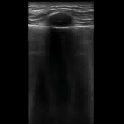
user: Please summarize the details of <|img1|>.
assistant:  An ultrasound image depicting lung using a linear probe. This shows clear signs of Pneumonia. This image reflects a LUSS of 1. This is likely a soft tissue ultrasound using a linear transducer in B-mode, typically employed for superficial structures.

Visible Organs and Structures: The image predominantly shows subcutaneous tissue and possibly a mass or cystic structure near the surface.

Features of Each Organ/Structure:
- Size: The structure appears slightly raised or protruding, suggesting it could be either normal or slightly enlarged.
- Shape: It appears to have a rounded shape, which is typically consistent with a cystic lesion.
- Texture (echotexture): The structure is predominantly anechoic, indicating fluid content, which aligns with cystic characteristics.
- Borders: The borders of the structure are well-defined, typical of fluid-filled cysts.
- Symmetry: Symmetrical as a singular observable structure.

Additional Findings:
- Fluid Presence: The anechoic nature suggests a clear fluid collection within the structure, likely indicating a cyst.
- Artifacts: There might be posterior acoustic enhancement, a common feature with fluid-filled structures.
- Masses/Lesions: The appearance suggests a simple cyst based on the anechoic interior and defined borders.
- Vascular Features: Not applicable as no Doppler mode is used.

Gray Scale and Doppler Features: Hypoechoic to anechoic regions indicate a cystic structure; without Doppler data, no vascular flow pattern can be determined.

Dynamic Features: Not applicable in a static image assessment without motion visuals.

Image Quality and Limitations: The image quality appears to be good, giving clear structural details. Limitations include the inability to assess vascular features or deeper structures without further imaging or Doppler use.

Patient Demographic Context: Not available from the given image.

Impression/Conclusion: The ultrasound image likely indicates a simple cyst within the soft tissue, given the anechoic characteristics and defined borders. No complicating features such as solid components or irregular borders are present, suggesting a benign process. Further clinical correlation and possibly Doppler imaging would help confirm diagnosis.
|
21
|
covid_us__00000020
|
|
user: What can you say about this image: <|img1|>?
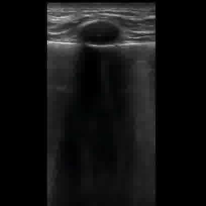
assistant:  An ultrasound image depicting lung using a linear probe. The patient shows symptoms of Pneumonia. The computed LUSS is 1. This image appears to be from a soft tissue ultrasound, likely involving a linear transducer in B-mode. It is typically used to evaluate superficial structures.

Visible Organs and Structures: The image shows a superficial tissue with a dark, round structure indicative of a cyst or a lesion located near the surface of the skin.

Features of Each Organ/Structure:
- Size: The round structure could be measured for precise dimensions, though not available from this image alone.
- Shape: The structure is round and appears well-defined.
- Texture (echotexture): The structure is anechoic, suggesting fluid content.
- Borders: Smooth borders suggest it is well-encapsulated.
- Symmetry: Not applicable for this singular structure.

Additional Findings:
- Fluid Presence: The anechoic nature of the structure indicates a possible fluid-filled cyst.
- Artifacts: Posterior acoustic enhancement is noted, common with fluid-filled structures.
- Masses/Lesions: The structure is consistent with a simple cyst based on its echotexture and posterior enhancement.
- Vascular Features: Not applicable as Doppler mode is not visible in the image.

Gray Scale and Doppler Features: Hypoechoic regions suggest soft tissue surrounding the anechoic cyst. Doppler not applicable.

Dynamic Features: No dynamic features can be assessed from this static image.

Image Quality and Limitations: The image quality appears good, with clear visualization of the anechoic structure. No significant limitations noted; however, clinical correlation with patient history and symptoms is necessary.

Patient Demographic Context: Patient age and sex are not available.

Impression/Conclusion: The ultrasound image reveals a well-defined, anechoic structure with posterior acoustic enhancement consistent with a simple cyst in the superficial tissues. Further clinical assessment is recommended to confirm the diagnosis and guide management. If symptomatic or atypical features are present, further evaluation may be necessary.
|
22
|
covid_us__00000021
|
|
user: What can be observed in <|img1|>?
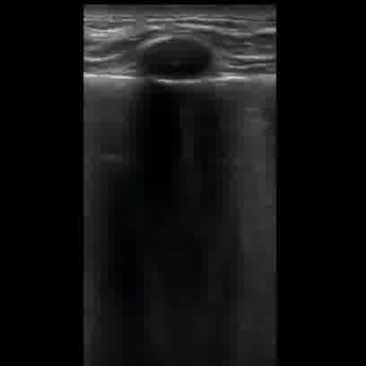
assistant:  Ultrasound imaging of lung performed with a linear probe. Signs of Pneumonia are evident in this image. LUSS is measured at 1. This image appears to be a soft tissue ultrasound using a linear transducer, most likely captured in B-mode.

Visible Organs and Structures: The ultrasound image predominantly displays superficial soft tissue structures. It includes visible layers typical of skin and subcutaneous tissues.

Features of Each Organ/Structure:
- Visible Structures: The skin and subcutaneous tissues are well-visualized. The image shows an anechoic structure beneath the skin, likely suggesting a cystic lesion.
 - Size: The anechoic structure appears round and localized. Exact measurements aren't possible from the image alone.
 - Shape: The structure is round with a well-defined border.
 - Texture: The lesion is anechoic, consistent with fluid content.
 - Borders: Borders are smooth and well-defined.

Additional Findings:
- Fluid Presence: The anechoic area suggests a fluid-filled cyst or lesion beneath the skin.
- Artifacts: Posterior acoustic enhancement is present, reinforcing the fluid-filled nature of the structure.
- Masses/Lesions: A probable simple cyst is present, given the anechoic appearance and acoustic enhancement.
- Vascular Features: No Doppler features or vascular flow information available in this image.

Gray Scale and Doppler Features: The anechoic region is clinically significant for indicating a fluid collection, likely a simple cyst. No Doppler data available.

Dynamic Features: Not applicable in this static image.

Image Quality and Limitations: The image quality is good, leading to clear visualization of structures. Limitations include the absence of Doppler and motion assessment.

Patient Demographic Context: Patient age and sex are not provided.

Impression/Conclusion: The ultrasound image reveals an anechoic, well-circumscribed lesion in the subcutaneous tissue, suggestive of a simple cyst. Further clinical correlation and possibly additional imaging or aspiration may be needed for definitive diagnosis.
|
23
|
covid_us__00000022
|
|
user: What can be observed in <|img1|>?
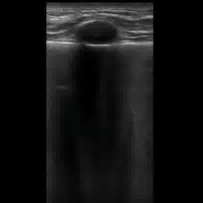
assistant:  This ultrasound highlights lung with the help of a linear probe. The patient is diagnosed with Pneumonia. This image reflects a LUSS of 1. This is an ultrasound image using a linear transducer, likely in B-mode, which is often used for soft tissue evaluation.

Visible Organs and Structures: The image primarily shows layers of soft tissue. The top layers may represent skin and subcutaneous tissue, with a defined structure visible just below the surface.

Features of Each Organ/Structure:
- Size: The visible structure appears as a small, focal lesion.
- Shape: The lesion is oval and well-defined.
- Texture (echotexture): Appears hypoechoic compared to surrounding tissue.
- Borders: The borders of the lesion are smooth and well-defined.
- Symmetry: No bilateral structures to compare in this image.

Additional Findings:
- Fluid Presence: There is no evident fluid collection around the lesion.
- Artifacts: There might be shadowing beneath the lesion, suggesting a solid structure.
- Masses/Lesions: The lesion is a small, well-defined hypoechoic mass, likely benign.
- Vascular Features: No vascular features visible in this static image.

Gray Scale and Doppler Features: The central hypoechoic region may indicate a cystic or solid nature, common in benign lesions. No Doppler information on flow is provided.

Dynamic Features: Not applicable, as a static image is presented.

Image Quality and Limitations: The image quality is good; however, interpretation is limited by the lack of dynamic information and Doppler data.

Patient Demographic Context: No specific information on the patient's age or sex is provided.

Impression/Conclusion: The ultrasound image reveals a well-defined, oval, hypoechoic structure in the soft tissue, likely indicating a benign lesion such as a cyst or lipoma. Further assessment with Doppler ultrasound could help in evaluating any vascularity within the lesion.
|
24
|
covid_us__00000023
|
|
user: Describe what you see in <|img1|>.
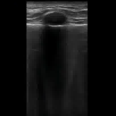
assistant:  Ultrasound imaging of lung performed with a linear probe. The patient is suffering from Pneumonia. LUSS is measured at 1. This is a musculoskeletal ultrasound using a linear transducer in B-mode.

Visible Organs and Structures: The image primarily shows subcutaneous tissue and possibly junction with deeper structures, as typical in soft tissue evaluations.

Features of Each Organ/Structure:
- Size and Shape: The structure appears to be regular in shape.
- Texture (echotexture): There is a well-defined, anechoic area suggesting a fluid collection.
- Borders: The borders of the anechoic area are smooth.
- Symmetry: Symmetry is not applicable as this is a localized view.

Additional Findings:
- Fluid Presence: An anechoic area indicates a well-circumscribed fluid collection, possibly a cyst or seroma.
- Artifacts: Acoustic enhancement is present, common with fluid collections. There may be shadowing at the edges due to the density difference between fluid and surrounding tissue.

Gray Scale and Doppler Features: Hypoechoic to anechoic regions are clinically significant for fluid presence. Doppler was not used in this image, so vascular flow information is not available.

Dynamic Features: Not applicable as this is a static image.

Image Quality and Limitations: Image quality is good, but limitations include a lack of Doppler information and context of surrounding anatomical landmarks for more comprehensive evaluation.

Patient Demographic Context: Patient age and sex are not provided and could influence interpretation (e.g., distinguishing between types of cysts).

Impression/Conclusion: The image likely demonstrates a simple cyst or fluid collection given the anechoic region with smooth borders and posterior acoustic enhancement. Further clinical correlation and history are advised for accurate diagnosis.
|
25
|
covid_us__00000024
|
|
user: Can you describe <|img1|> in detail?
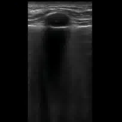
assistant:  A linear probe captured this ultrasound of lung. The findings indicate the presence of Pneumonia. The LUSS score is 1. This ultrasound image likely represents a soft tissue neck ultrasound using a linear transducer in B-mode.

Visible Organs and Structures: The image focuses on superficial soft tissues, with the dermis and subcutaneous layers being well-visualized.

Features of Each Structure:
- Subcutaneous Tissue:
 - Size: Appears normal; measurements are not specified.
 - Shape: Regular.
 - Texture (echotexture): Relatively homogeneous.
 - Borders: Smooth.
 - Symmetry: Not applicable for a single-plane view.

Additional Findings:
- Fluid Presence: A potential anechoic or hypoechoic area is seen, which might represent a cystic structure.
- Artifacts: Prominent acoustic shadowing beneath the area suggests anterior acoustic enhancement, typical of cystic structures.

Gray Scale and Doppler Features:
- The hyperechoic and anechoic areas may indicate a normal fat lobule with shadowing or a cystic lesion. Doppler was likely not used here as vascular flow isn't assessed.

Dynamic Features:
- Static image; dynamic features such as motion or changes with respiration are not applicable.

Image Quality and Limitations:
- Image quality appears fair, although deeper structures are obscured by shadowing. The linear probe offers detailed superficial views but limited penetration for deeper tissues.

Patient Demographic Context:
- No specific patient age or sex is available.

Impression/Conclusion:
- The image most likely demonstrates a soft tissue structure with a cystic characteristic, given the anechoic center and posterior acoustic enhancement. If clinical correlation and symptoms suggest, further evaluation might be needed to confirm a diagnosis such as a sebaceous cyst or other similar lesions. Evaluation with Doppler could help rule out vascular lesions or anomalies.
|
26
|
covid_us__00000025
|
|
user: How would you summarize <|img1|> visually?
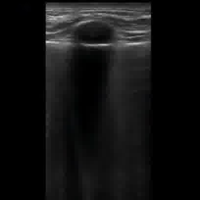
assistant:  An ultrasound image depicting lung using a linear probe. The findings indicate the presence of Pneumonia. A LUSS of 1 is reported. This image appears to be obtained using a high-frequency linear transducer in B-mode, typical for superficial structures, possibly musculoskeletal or soft tissue ultrasound.

Visible Organs and Structures: The image shows superficial soft tissues. Deeper structures are not visible, likely due to the focal area of interest and the limitations of the transducer used.

Features of Each Organ/Structure:

- Size: Not applicable as specific structures are not identified.
- Shape: Not applicable; focus seems to be on soft tissue.
- Texture (echotexture): Predominantly anechoic region in the central portion suggests fluid presence or a cystic structure.
- Borders: Clear, smooth borders are evident in the anechoic region.
- Symmetry: Not applicable due to no bilateral structures being visible.

Additional Findings:

- Fluid Presence: Anechoic area suggests fluid collection or a cystic structure.
- Artifacts: Shadowing from a superficial mass or lesion, indicative of a possible solid structure or foreign body causing acoustic shadowing.
- Masses/Lesions: Potential presence of a well-defined lesion causing posterior shadowing.
- Vascular Features: Not assessed in this mode.

Gray Scale and Doppler Features: Clinical significance of an anechoic region could indicate a fluid-filled structure or cyst. No Doppler features are assessed here.

Dynamic Features: N/A, no real-time assessment provided.

Image Quality and Limitations: Image quality appears fair but restricted by depth penetration of the transducer. Overlying soft tissue may be obscuring deeper structures.

Patient Demographic Context: Not provided; helps narrow differential diagnosis.

Impression/Conclusion: The ultrasound image suggests the presence of an anechoic cystic area in the superficial tissues with posterior shadowing, possibly indicative of a cyst, fluid accumulation, or foreign body. Further evaluation or clinical correlation is needed for a definitive diagnosis.
|
27
|
covid_us__00000026
|
|
user: Here is an image: <|img1|>. Describe it
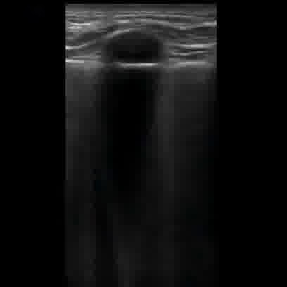
assistant:  Ultrasound imaging of lung performed with a linear probe. The patient is suffering from Pneumonia. LUSS scoring indicates a value of 1. This is a soft tissue ultrasound using a linear transducer, likely in B-mode. It seems focused on a region near the skin surface.

Visible Organs and Structures: The primary structure is a cystic lesion located in the subcutaneous tissue. The overlying skin and superficial tissues are well visualized.

Features of Each Organ/Structure:
- Size: The lesion appears small to moderately sized. Exact measurements are not provided.
- Shape: The lesion is round with a smooth contour.
- Texture (echotexture): The lesion is anechoic with posterior acoustic enhancement, indicating fluid content.
- Borders: The borders are well-defined and smooth.
- Symmetry: Not applicable as there's a singular focus.

Additional Findings:
- Fluid Presence: There is a clear anechoic area suggesting a fluid-filled cyst.
- Artifacts: Presence of posterior acoustic enhancement is noted, common with cystic structures.
- Masses/Lesions: A single well-defined cystic lesion as described.
- Vascular Features: No vascular features are evident within the lesion; Doppler may not have been used.

Gray Scale and Doppler Features: The cyst is anechoic, consistent with a simple fluid-filled structure.

Dynamic Features: No motion features noted, considering it's a static soft tissue image.

Image Quality and Limitations: The image quality appears good for assessing superficial lesions. No significant artifacts impact the interpretation. Deeper structures are poorly visualized with this view, likely by design.

Patient Demographic Context: No specific patient age or sex provided.

Impression/Conclusion: The ultrasound image demonstrates a well-defined anechoic cystic lesion in the subcutaneous tissue, likely a benign simple cyst or sebaceous cyst. Clinical correlation and possibly further imaging could help confirm the diagnosis if any intervention is planned.
|
28
|
covid_us__00000027
|
|
user: What can you say about this image: <|img1|>?
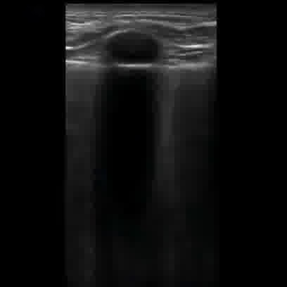
assistant:  This ultrasound highlights lung with the help of a linear probe. The patient shows symptoms of Pneumonia. LUSS is measured at 1. This appears to be a soft tissue ultrasound using a linear transducer in B-mode.

Visible Organs and Structures: The image shows superficial tissue with a well-circumscribed structure suggestive of a cyst or lesion. Deeper tissues are not clearly visualized due to shadowing.

Features of Each Organ/Structure:
- Size: The structure appears round and is likely enlarged for the expected visibility at this depth.
- Shape: The structure is round and regular.
- Texture (echotexture): The structure is predominantly anechoic, indicating fluid content.
- Borders: The borders are smooth and well-defined.
- Symmetry: Not applicable as this is a single structure view.

Additional Findings:
- Fluid Presence: The anechoic nature suggests a fluid-filled cyst.
- Artifacts: Notable posterior acoustic shadowing below the structure, consistent with its fluid-filled nature.
- Masses/Lesions: The lesion is likely a simple cyst given its anechoic center and posterior enhancement.
- Vascular Features: No Doppler vascular information is visible in this image.

Gray Scale and Doppler Features: The anechoic region likely correlates with a benign fluid-filled cyst. Doppler was not applied here.

Dynamic Features: N/A for this static image.

Image Quality and Limitations: The image quality is good but limited by posterior shadowing, preventing visualization of deeper structures.

Patient Demographic Context: Age and sex not available.

Impression/Conclusion: The ultrasound image demonstrates a well-defined anechoic structure with posterior acoustic enhancement, likely indicating a benign cyst. Further evaluation with Doppler and clinical correlation is recommended for comprehensive assessment.
|
29
|
covid_us__00000028
|
|
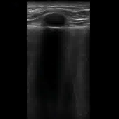
user: What can you say about this image: <|img1|>?
assistant:  This is an image of lung obtained using a linear probe. The results are consistent with a diagnosis of Pneumonia. The LUSS score is 1. This is an image from a soft tissue ultrasound, likely using a linear transducer in B-mode.

Visible Organs and Structures: The ultrasound image shows a superficial soft tissue mass. The surrounding soft tissue is visible, but underlying structures are not well visualized, likely due to the nature of the mass and its shadowing effect.

Features of Each Organ/Structure:
- Mass:
 - Size: Appears medium in size; precise measurements are not provided.
 - Shape: The mass is oval and regular in shape.
 - Texture (echotexture): The mass is hyperechoic (bright) with posterior acoustic shadowing.
 - Borders: The mass has well-defined borders.
 - Symmetry: Not applicable as this is a singular lesion.

Additional Findings:
- Fluid Presence: No significant fluid collections are visible.
- Artifacts: Notable shadowing artifact deep to the mass is present.
- Masses/Lesions: The principal finding is the hyperechoic mass with posterior shadowing.
- Vascular Features: Not evaluated as no Doppler mode is mentioned in the image.

Gray Scale and Doppler Features:
- The hyperechoic region suggests the presence of a calcified or dense lesion.
- Doppler evaluation is not provided, so flow characteristics are not assessed.

Dynamic Features:
- Organ motion and valvular motion are not applicable in this static image.

Image Quality and Limitations:
- Image quality appears good for superficial evaluation but limited for deeper structures due to shadowing. Soft tissue positioning seems adequate for the target lesion.

Patient Demographic Context: Information not provided.

Impression/Conclusion: The ultrasound shows a well-defined, hyperechoic mass with posterior shadowing in the soft tissue, suggestive of a calcified lesion or foreign body. Further clinical correlation and possibly additional imaging may be needed for a definitive diagnosis.
|
30
|
covid_us__00000029
|
|
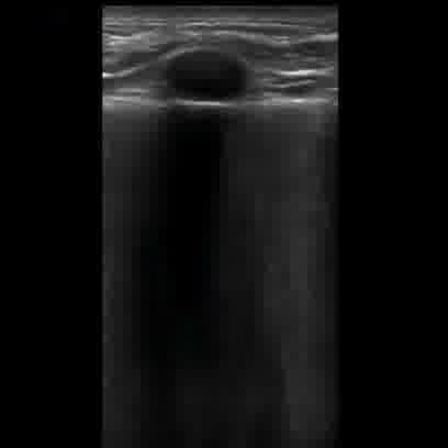
user: What can you say about this image: <|img1|>?
assistant:  This is an image of lung obtained using a linear probe. The findings indicate the presence of Pneumonia. A LUSS of 1 is reported. This appears to be an ultrasound of the thyroid. A linear transducer is likely used, operating in B-mode.

Visible Organs and Structures: The thyroid gland is visible, along with overlying subcutaneous tissue and skin. Only a portion of the thyroid is well-visualized in this image.

Features of Each Organ/Structure:
- Thyroid:
 - Size: Normal size for the part visualized.
 - Shape: Regular contour of the visible portion.
 - Texture (echotexture): The part of the thyroid seen appears heterogeneous.
 - Borders: Smooth borders where visible.
 - Symmetry: Only one side is visible, preventing a bilateral comparison.

Additional Findings:
- Fluid Presence: No fluid collections apparent in this image.
- Artifacts: There is significant acoustic shadowing beneath the structure, possibly indicating a cystic area.
- Masses/Lesions: A well-defined, anechoic lesion is present, likely a benign cyst based on echotexture and shadowing.
- Vascular Features: Not assessed in this B-mode image.

Gray Scale and Doppler Features: The anechoic area is likely a cyst, indicating a non-solid lesion. No Doppler analysis provided in this image.

Dynamic Features: No dynamic assessment applicable as this is a static image.

Image Quality and Limitations: The image quality appears good, with some shadow artifact affecting deeper visualization. There may be limitations in assessing the entire thyroid due to positioning.

Patient Demographic Context: No age or sex information provided.

Impression/Conclusion: The ultrasound image shows a well-defined anechoic lesion in the thyroid, consistent with a simple cyst. No suspicious features suggesting malignancy are identified in this limited view. Further evaluation with full thyroid ultrasound and potential Doppler study may be warranted if clinically indicated.
|
31
|
covid_us__00000030
|
|
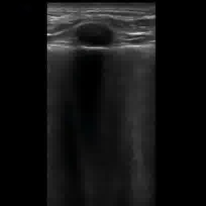
user: How would you summarize <|img1|> visually?
assistant:  Ultrasound imaging of lung performed with a linear probe. Signs of Pneumonia are evident in this image. The computed LUSS is 1. Likely a soft tissue ultrasound using a linear transducer, typically in B-mode.

Visible Organs and Structures: Primarily subcutaneous tissues and a well-defined lesion.

Features of Each Organ/Structure:
- Lesion:
 - Size: Appears medium-sized; exact measurements not available.
 - Shape: Round.
 - Texture (echotexture): Anechoic center suggestive of fluid or cystic nature.
 - Borders: Well-defined and smooth.
 - Symmetry: Not applicable as it is a singular lesion.

Additional Findings:
- Fluid Presence: Anechoic nature suggests fluid content, potentially a cyst.
- Artifacts: Posterior acoustic enhancement is visible indicating the fluid nature of the lesion.
- Masses/Lesions: The lesion's appearance is consistent with a simple cyst.
- Vascular Features: Not applicable as Doppler mode was not mentioned.

Gray Scale and Doppler Features: The anechoic region indicates probable fluid content. Doppler findings not applicable.

Dynamic Features: No significant organ motion noted as this is a static soft tissue structure.

Image Quality and Limitations: Image quality is adequate for identifying the lesion, though without Doppler features or dynamic assessment. Limited field suggests soft tissue rather than deep organ involvement.

Patient Demographic Context: Not provided; typically affects a wide demographic.

Impression/Conclusion: The image suggests a well-circumscribed, anechoic lesion consistent with a simple cyst in the soft tissue. Further assessment, potentially with Doppler or follow-up imaging, may be advised to confirm diagnosis. No evidence of malignancy seen based on the current image.
|
32
|
covid_us__00000031
|
|
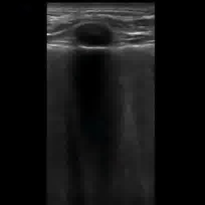
user: Here is an image: <|img1|>. Describe it
assistant:  This ultrasound image shows lung using a linear probe. The patient is diagnosed with Pneumonia. The calculated LUSS value is 1. This is an ultrasound image likely obtained using a linear transducer. The mode appears to be B-mode, which is typical for viewing superficial structures.

Visible Organs and Structures: The image primarily shows subcutaneous tissue and possibly the upper part of a limb. There is a distinct round structure at the top, likely a transverse view of a cyst or lesion.

Features of Each Organ/Structure:
- Round Structure:
 - Size: Appears as small to moderate; measurements are not provided.
 - Shape: Regular and round.
 - Texture (echotexture): Anechoic, indicating fluid content.
 - Borders: Smooth and well-defined.
 - Symmetry: N/A as it is a single structure.

Additional Findings:
- Fluid Presence: The round structure is anechoic, suggesting a fluid collection like a cyst.
- Artifacts: There is posterior acoustic enhancement beneath the anechoic area, typical for fluid-filled structures.
- Masses/Lesions: The round anechoic area could indicate a benign cystic lesion based on its appearance.
- Vascular Features: No vascular structures are visible in this still image.

Gray Scale and Doppler Features: The anechoic region suggests a fluid-filled space. No Doppler information is available in this image to assess any flow characteristics.

Dynamic Features: Not assessable in a still image.

Image Quality and Limitations: Image quality appears satisfactory, with good definition of structures. There might be some limitations due to the image being still and not showing depth or effects with compression.

Patient Demographic Context: Not available; it is important to consider patient age and sex for further clinical correlation.

Impression/Conclusion: The image suggests a well-defined anechoic structure consistent with a simple cyst or similar benign fluid collection. Clinical correlation and further imaging, potentially with Doppler, might help in differentiating if vascularization or other features are noted.
|