| # | key | img1.png | txt |
|---|
1
|
medtrinity_conv_2__00000000
|
|
user: Summarize the content of the image as if it were an alternative text (alt text) for accessibility. <|img1|>
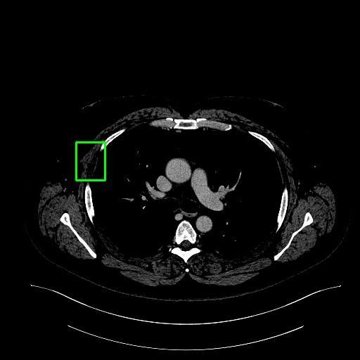
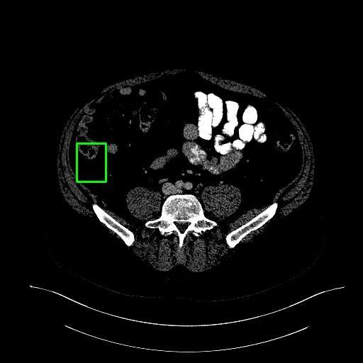
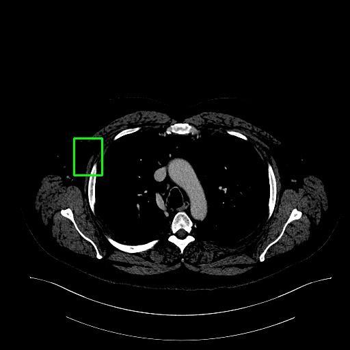
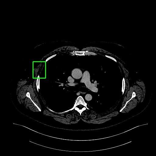
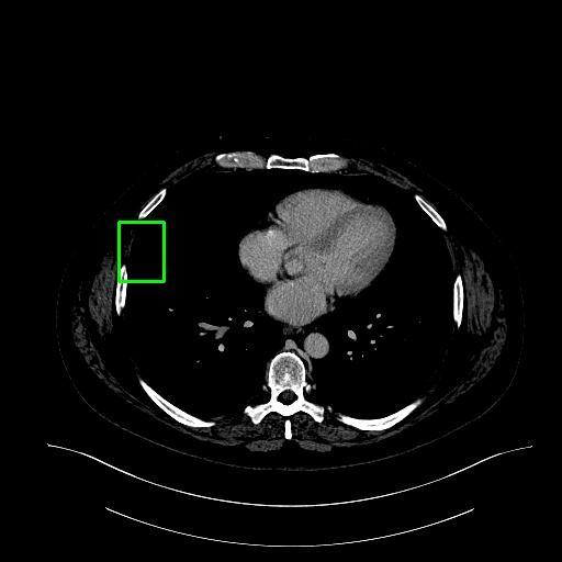
assistant:  This CT scan image highlights the liver, with a specific focus on a lesion located in the left-center horizontally and the middle vertically. The lesion occupies approximately 0.8% of the total area.

Key observations:
- The lesion exhibits an unusual appearance, differing from the surrounding hepatic tissue in color, texture, or size.
- These characteristics suggest a potential liver lesion.
- Its localized nature may indicate a focal pathological process.
- While it does not appear to directly affect adjacent tissue, it could be related to or contribute to an underlying hepatic disease state.
|
2
|
medtrinity_conv_2__00000001
|
|
user: Describe everything visible in this image in a clear, factual way. <|img1|>
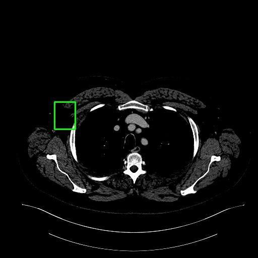
assistant:  The image is a computed tomography (CT) scan showing a cross-sectional view of the upper abdomen, with the liver prominently visible in the left-center region.

Key observations:
- The liver occupies a large portion of the upper abdomen.
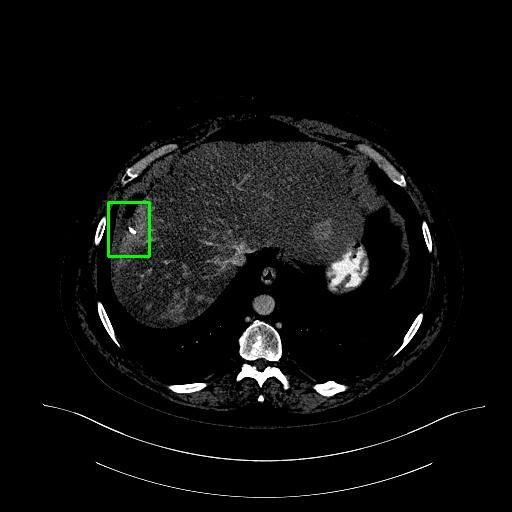
- Other structures, such as the lungs and parts of the gastrointestinal tract, are also visible.
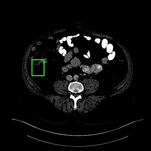
- A region of interest is located horizontally in the left-center and vertically in the middle of the image, covering approximately 0.8% of the total area.
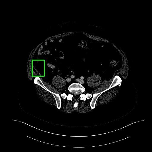
- This region exhibits an unusual appearance compared to the surrounding liver tissue, characterized by distinct differences in density or texture.
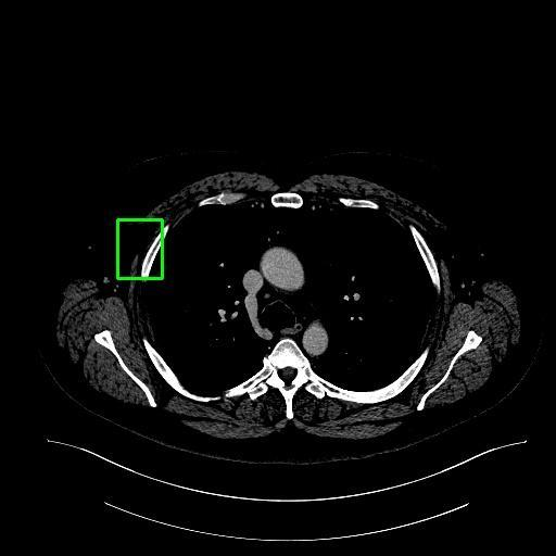
- These characteristics suggest the presence of a potential lesion within the liver.

Implications:
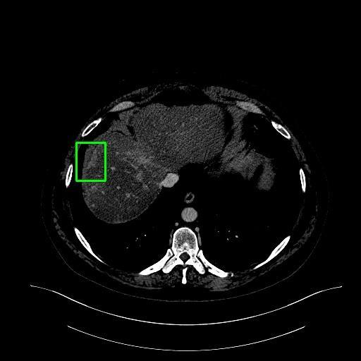
- The lesion appears localized and may indicate a pathological change.
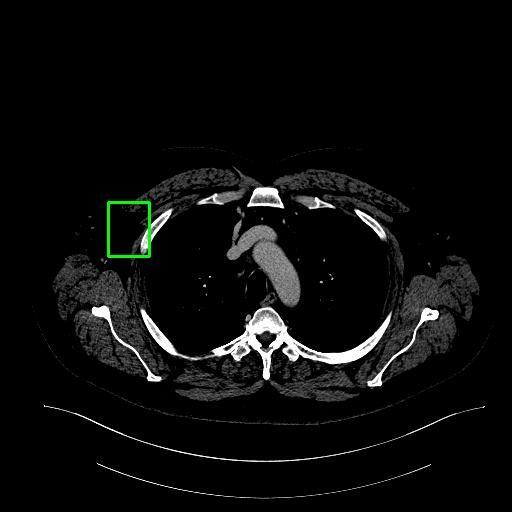
- Its proximity to healthy liver tissue suggests it could be affecting—or being affected by—adjacent hepatic structures.
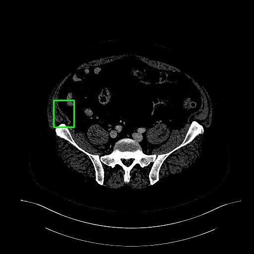
|
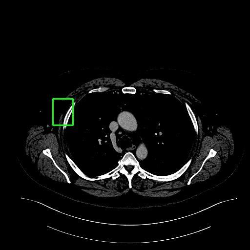
3
|
medtrinity_conv_2__00000002
|
|
user: Provide a concise description of this image, focusing on the main subject and background. <|img1|>
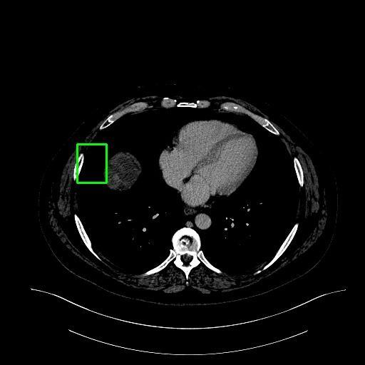
assistant:  The image is a transverse CT scan of the abdomen, with a focus on the liver.

- The liver is the dominant organ visible in the scan, primarily located on the right side of the abdomen but extending across the midline to the left.
- A region of interest is identified in the left-center portion of the liver, occupying approximately 0.8% of the total area.
- This area exhibits an abnormality in texture and possibly density compared to the surrounding liver parenchyma, suggesting the presence of a lesion.
- The lesion’s location may indicate a potential relationship with nearby liver structures, such as vascular pathways or other anatomical features.
- It could represent a localized manifestation of a broader liver condition or may be influencing adjacent structures.

Further clinical evaluation and additional diagnostic information are required to determine the exact nature and significance of this finding.
|
4
|
medtrinity_conv_2__00000003
|
|
user: Provide a concise description of this image, focusing on the main subject and background. <|img1|>
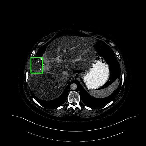
assistant:  The image is a computed tomography (CT) scan of the abdomen, with a focus on the liver.

### Liver Location and Appearance
- The liver is situated in the **upper right quadrant** of the abdomen, just below the diaphragm.
- It appears as a large, dark region in the scan, with other abdominal structures visible in the surrounding area.

### Abnormality Detected
- A region of interest is located **left-center and vertically centered** within the liver.
- This area represents **0.8%** of the total image area and exhibits an abnormality in liver tissue.
- The abnormality is characterized by:
  - Altered **texture**
  - Different **density** compared to the surrounding liver parenchyma

### Clinical Implications
- The findings suggest the presence of a **localized liver lesion**.
- Its proximity to healthy liver tissue may indicate:
  - A condition confined to a specific region
  - Potential interaction with or impact on adjacent hepatic structures
- Given the liver’s functional unity, such lesions may reflect or influence overall liver function, as liver diseases often affect broader organ systems.
|
5
|
medtrinity_conv_2__00000004
|
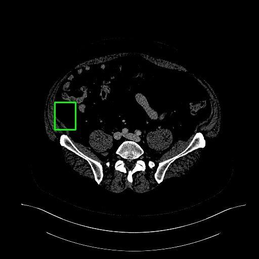
|
user: Create an image caption that would help someone understand the picture without seeing it. <|img1|>
assistant:  The image is a computed tomography (CT) scan of the abdomen, with a focus on the liver.

### Liver Location and Appearance
- The liver is situated in the **upper right quadrant** of the abdomen, just below the diaphragm.
- It appears as a large, dark region in the scan, with surrounding abdominal organs clearly visible.

### Abnormality Detected
- A region of interest is located **left-center and vertically centered** within the liver.
- This area represents **0.8%** of the total liver region.
- The abnormality is characterized by:
  - A noticeable difference in **texture**.
  - A possible variation in **density** compared to the surrounding liver parenchyma.

### Clinical Implication
- These features suggest the presence of a **liver lesion**.
- The lesion’s proximity to healthy liver tissue may indicate:
  - A **localized manifestation** of a broader hepatic condition.
  - Potential **involvement of adjacent liver tissue**.

Further evaluation is recommended to determine the nature and cause of this abnormality.
|
6
|
medtrinity_conv_2__00000005
|
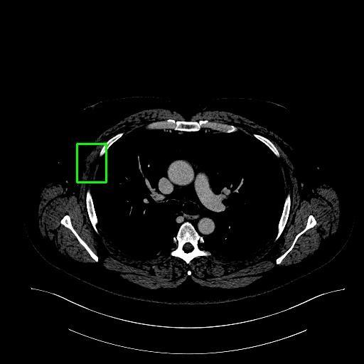
|
user: Generate a caption that accurately represents the content of this image. <|img1|>
assistant:  The image is a transverse CT scan of the abdomen, with a focus on the liver.

- The liver is prominently visible in the upper portion of the image.
- The heart and portions of the rib cage are also present.

A region of interest is located in the left-center to middle of the image, showing an area within the liver with a distinct texture and density compared to the surrounding liver tissue. This difference suggests the presence of a **focal liver lesion**.

Possible causes of this lesion include:
- Tumor (benign or malignant)
- Cyst
- Abscess

The lesion is situated within the liver parenchyma and may reflect:
- A localized abnormality
- A manifestation of a systemic condition affecting the liver
- A diffuse liver disease process that has produced focal changes

Further clinical and imaging evaluation is warranted to determine the exact nature and implications of this finding for liver function.
|
7
|
medtrinity_conv_2__00000006
|
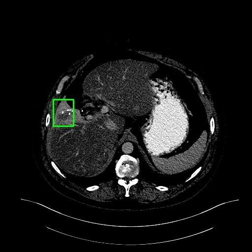
|
user: Explain what is shown in the image. <|img1|>
assistant:  The image is a transverse CT scan of the abdomen, with a focus on the liver.

- The liver is the dominant organ visible in the scan, primarily located on the right side of the abdomen but extending across the midline to the left.
- A region of interest is identified in the left-center portion of the liver, occupying approximately 0.8% of the total area.
- This area exhibits an abnormality in texture and possibly density compared to the surrounding liver parenchyma, suggesting the presence of a lesion.
- The lesion’s location may indicate a potential relationship with nearby liver structures, such as:
  - A localized manifestation of a broader liver condition.
  - An impact on adjacent vascular structures due to its presence.
- Further clinical evaluation and additional diagnostic information are required to determine the exact nature and significance of this finding.
|
8
|
medtrinity_conv_2__00000007
|
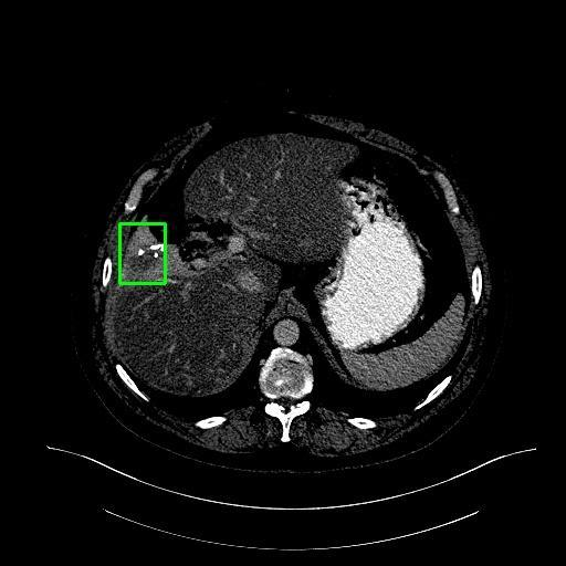
|
user: What do you see in this image? <|img1|> Describe it in detail.
assistant:  The image is a transverse CT scan of the abdomen, with a focus on the liver.

- The liver occupies a large portion of the upper abdomen, primarily on the right side.
- A region of interest is identified in the left-center, vertically centered, accounting for 0.8% of the total area.
- This region exhibits an abnormal texture and/or density compared to the surrounding liver parenchyma, suggesting a possible lesion.
  - Potential causes include:
    - Tumor
    - Cyst
    - Fatty infiltration

The lesion’s location within the liver and its proximity to other visible structures—such as the heart and lungs—may indicate interactions with adjacent hepatic tissue. Its characteristics and spatial relationship to the surrounding liver could point to a localized hepatic condition, potentially affecting overall liver function or health.
|
9
|
medtrinity_conv_2__00000008
|
|
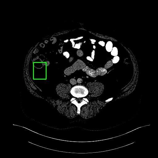
user: Create an accurate image description for a machine learning dataset, covering all key elements. <|img1|>
assistant:  The image is a coronal CT scan of the abdomen, with a focus on the liver.

### Key Observations:
- The **liver** is prominently visible in the upper portion of the image.
- Other abdominal structures are also present, including:
  - The **spleen**
  - The **kidneys**
  - Segments of the **bowel**

### Lesion Details:
- A distinct **lesion** is located in the left-center to middle region of the liver.
- It appears **darker** than the surrounding liver tissue, indicating a difference in density and texture.
- This hypodense appearance suggests possible causes such as:
  - A **cyst**
  - An **abscess**
  - A **neoplastic process** (e.g., benign or malignant tumor)

### Clinical Implications:
- The lesion is in close proximity to healthy liver parenchyma, suggesting potential interaction with surrounding tissue.
- Its presence may reflect or contribute to altered liver function.
- It could be a primary liver abnormality or a manifestation of an underlying systemic condition.

Further evaluation is recommended to determine the exact nature of the lesion.
|
10
|
medtrinity_conv_2__00000009
|
|
user: What do you see in this image? <|img1|> Describe it in detail.
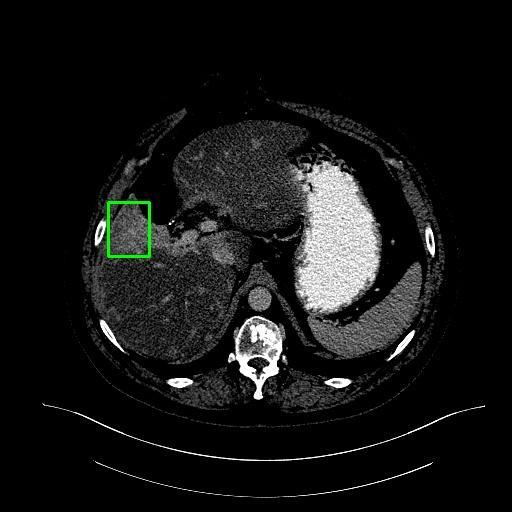
assistant:  The image is a transverse CT scan of the abdomen, with a focus on the liver.

- The liver is the dominant organ visible in the scan, primarily located on the right side of the abdomen but extending across the midline to the left.
- A region of interest, situated in the left-center of the image, occupies approximately 0.8% of the total area and exhibits an abnormality within the liver tissue.
- This abnormality appears as a distinct change in texture and/or density compared to the surrounding liver parenchyma, suggesting the presence of a lesion.
- Due to its location, the lesion may be in close proximity to other liver structures, raising the possibility of a relationship—such as a localized manifestation of a broader hepatic condition or an impact on nearby vascular structures.
- The precise nature of this lesion and its clinical significance requires further evaluation through additional diagnostic tests and clinical correlation.
|
11
|
medtrinity_conv_2__00000010
|
|
user: Provide an objective description of what appears in the image. <|img1|>
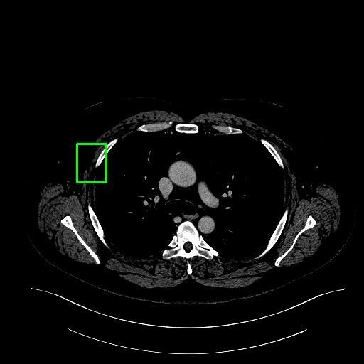
assistant:  The image is a computed tomography (CT) scan showing a cross-sectional view of the upper abdomen, with the liver prominently visible in the left-center region.

### Key Observations:
- The **liver** occupies a large portion of the upper abdomen.
- Other structures visible include the **lungs** and possibly parts of the **gastrointestinal tract**.
- A region of interest is located:
  - **Horizontally**: Left-center of the image
  - **Vertically**: Midline of the image
  - **Size**: Approximately 0.8% of the total image area

### Lesion Characteristics:
- This area exhibits an **unusual appearance** compared to the surrounding liver tissue.
- Differences may include:
  - **Density**
  - **Texture**
  - **Size**
- These features suggest a **potential pathological change** within the liver.

### Clinical Implications:
- The lesion’s proximity to other liver structures may indicate:
  - A **localized manifestation** of a broader liver condition.
  - Possible **interaction** with adjacent tissues—either being affected by them or affecting them in turn.

Further evaluation is recommended to determine the nature and significance of this finding.
|
12
|
medtrinity_conv_2__00000011
|
|
user: Summarize the content of the image as if it were an alternative text (alt text) for accessibility. <|img1|>
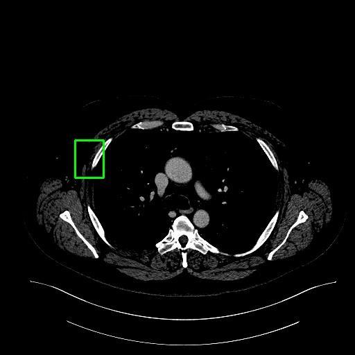
assistant:  The image is a transverse CT scan of the abdomen, with a focus on the liver.

- The liver is prominently visible in the upper portion of the image.
- Other abdominal organs, including the stomach and intestines, are also present.

A region of interest is located in the left-center to middle of the image, within the liver parenchyma. This area exhibits:

- A distinct texture and density compared to the surrounding liver tissue.
- Characteristics suggestive of a lesion.

This abnormality is likely the primary focus of the medical evaluation due to its clear contrast with normal hepatic tissue. Its localized nature indicates a potential pathological process that may involve or be influenced by adjacent liver structures.
|
13
|
medtrinity_conv_2__00000012
|
|
user: Generate a caption that identifies any visible text, signs, or labels in the image. <|img1|>
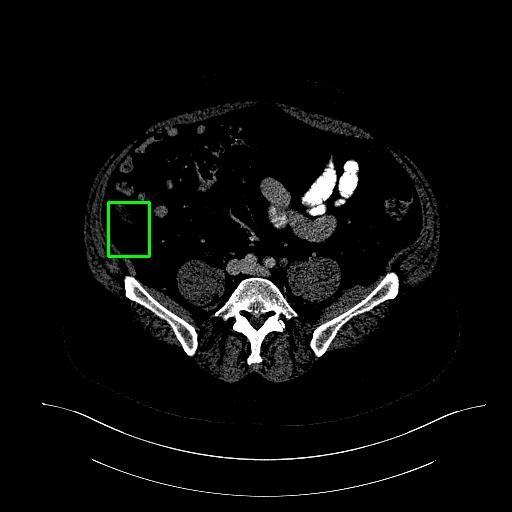
assistant:  The image is a coronal CT scan of the abdomen, with a focus on the liver.

### Key Observations:
- The **liver** is prominently visible in the upper portion of the image.
- Other abdominal structures are also present, including:
  - The **spleen**
  - The **kidneys**
  - Segments of the **bowel**

### Region of Interest:
- Located in the **left-center horizontally** and **mid-vertically**.
- Shows an area of **altered density** compared to the surrounding liver tissue.
- This discrepancy suggests a **potential lesion** within the liver.

### Clinical Implications:
- The lesion is distinct from normal liver parenchyma, indicating a possible **pathology**.
- Its location and appearance may suggest:
  - A **primary liver condition** (e.g., tumor, cyst, or abscess)
  - A **secondary manifestation** of a systemic disease

### Differential Considerations:
Based on imaging characteristics, the lesion could correspond to:
- **Benign or malignant tumors**
- **Cysts**
- **Abscesses**

Further evaluation is needed to determine the exact nature of the lesion.
|
14
|
medtrinity_conv_2__00000013
|
|
user: Create an accurate image description for a machine learning dataset, covering all key elements. <|img1|>
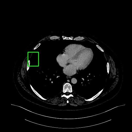
assistant:  The image is a transverse CT scan of the abdomen, with a focus on the liver.

- The liver is the dominant organ visible in the scan, primarily located on the right side of the abdomen but extending across the midline to the left.
- A region of interest, situated in the left-center of the image, occupies approximately 0.8% of the total area and exhibits an abnormality within the liver tissue.
- This abnormality appears as a distinct change in texture and/or density compared to the surrounding liver parenchyma, suggesting the presence of a lesion.
- Due to its location, the lesion may be in close proximity to other liver structures, potentially indicating:
  - A localized manifestation of a broader hepatic condition.
  - An impact on nearby vascular structures.

Further clinical evaluation and additional diagnostic information are required to determine the exact nature and significance of this finding.
|
15
|
medtrinity_conv_2__00000014
|
|
user: Generate a caption that identifies any visible text, signs, or labels in the image. <|img1|>
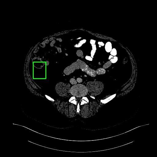
assistant:  The image is a coronal CT scan of the abdomen, with a focus on the liver.

### Key Observations:
- The **liver** is prominently visible in the upper portion of the image.
- Other abdominal structures are also present, including:
  - The **spleen**
  - The **kidneys**
  - Segments of the **bowel**

### Region of Interest:
- Located in the **left-center horizontally** and **mid-vertically**.
- Shows an area with **altered texture and density** compared to the surrounding liver tissue.
- This change is consistent with a **focal lesion**.

### Clinical Implications:
- The lesion’s distinct appearance suggests a **pathological process** within the liver.
- Its localized nature may indicate:
  - A **tumor** (benign or malignant)
  - A **cyst**
  - An **abscess**
- The lesion’s relationship with adjacent liver structures is critical for determining its origin and impact on liver function.

### Next Steps:
Further evaluation is recommended to confirm the nature of the lesion and assess its potential effect on overall hepatic health.
|
16
|
medtrinity_conv_2__00000015
|
|
user: Provide an objective description of what appears in the image. <|img1|>
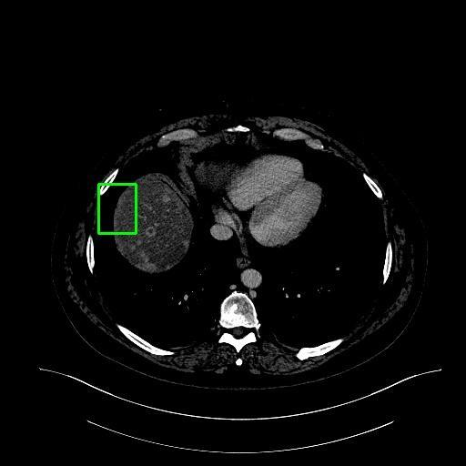
assistant:  This CT scan slice highlights the liver, with a notable lesion located in the left-center horizontally and the middle vertically. The lesion occupies approximately 0.8% of the total area.

Key observations:
- The lesion exhibits an unusual texture and density compared to the surrounding liver tissue, which may suggest a pathological change.
- Its proximity to adjacent liver structures indicates a potential interaction with the surrounding parenchyma.
- The lesion could be either a cause of localized liver changes or a manifestation of an underlying liver condition.

Further evaluation is recommended to determine the nature and clinical significance of this finding.
|
17
|
medtrinity_conv_2__00000016
|
|
user: Create an accurate image description for a machine learning dataset, covering all key elements. <|img1|>
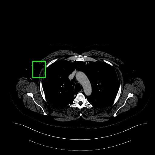
assistant:  This CT scan of the thoracic cavity shows the lungs clearly, with the heart positioned centrally between them.

Of particular interest is a distinct lesion in the liver, located in the left-center and mid-vertical region. Key observations include:

- The lesion has a different size and texture compared to the surrounding liver tissue.
- Its appearance is consistent with a nonmalignant process, such as a cyst or abscess.
- The lesion is localized, with clear proximity to adjacent hepatic parenchyma.

This suggests a focused process that may interact with nearby liver tissue, depending on the underlying cause. Further evaluation is recommended to confirm the nature of the lesion.
|
18
|
medtrinity_conv_2__00000017
|
|
user: Generate a caption that identifies any visible text, signs, or labels in the image. <|img1|>
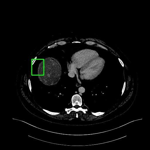
assistant:  The image is a transverse CT scan of the abdomen, with a focus on the liver.

- The liver is the dominant organ in the image, primarily located on the right side of the abdomen but extending across the midline to the left.
- A region of interest, situated in the left-center of the liver, occupies approximately 0.8% of the total area and exhibits an abnormality.
- This abnormality appears as a distinct change in texture and/or density compared to the surrounding liver parenchyma, suggesting the presence of a lesion.
- The lesion’s location may indicate a potential relationship with nearby liver structures, such as vascular pathways or adjacent tissue.
- Further clinical evaluation and additional diagnostic information are required to determine the exact nature of the lesion and its implications.
|
19
|
medtrinity_conv_2__00000018
|
|
user: Create an image caption that would help someone understand the picture without seeing it. <|img1|>
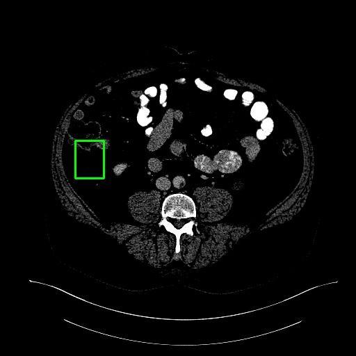
assistant:  The image is a coronal CT scan of the abdomen, with a focus on the liver.

- The liver is prominently visible in the upper portion of the image.
- Other abdominal structures, including the kidneys and segments of the intestines, are also present.

A region of interest is located in the **left-center horizontally** and **mid-vertically**, characterized by:

- **Altered density** compared to the surrounding liver parenchyma.
- A distinct appearance that suggests a possible **lesion** within the liver tissue.

This finding may indicate:

- A **primary liver condition**, such as a tumor, cyst, or abscess.
- A **secondary manifestation** of a systemic disease.

CT imaging is capable of detecting such abnormalities, and the altered density is consistent with known patterns of liver pathology. Further clinical correlation and imaging follow-up are recommended for definitive diagnosis.
|
20
|
medtrinity_conv_2__00000019
|
|
user: Generate a caption that identifies any visible text, signs, or labels in the image. <|img1|>
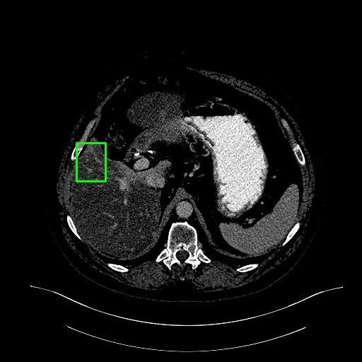
assistant:  The image is a transverse CT scan of the abdomen, with a primary focus on the liver.

### Liver Overview
- The liver is the largest organ visible in the scan, occupying most of the right upper quadrant and extending across the midline into the left side of the abdomen.

### Region of Interest
- A small abnormality is present in the left-center portion of the liver, covering approximately 0.8% of the total image area.
- This area exhibits a distinct difference in texture and/or density compared to the surrounding liver parenchyma.
- Potential causes include:
  - A tumor
  - A cyst
  - Fatty infiltration

### Anatomical Context
- The **spine** is centrally located and appears as a bright white, circular structure.
- The **abdominal wall** surrounds the abdominal cavity, providing clear anatomical boundaries.
- The lesion’s location suggests it may be interacting with or affected by adjacent hepatic structures.

### Clinical Implication
- The observed abnormality may indicate an underlying hepatic condition.
- It could potentially affect liver function, though further evaluation is needed to determine the exact nature and significance of the lesion.
|
21
|
medtrinity_conv_2__00000020
|
|
user: What do you see in this image? <|img1|> Describe it in detail.
assistant:  The image is a coronal CT scan of the abdomen, with a focus on the liver.

### Key Observations:
- The **liver** is prominently visible in the upper portion of the image.
- Other abdominal organs are also present, including the **spleen**, **kidneys**, and segments of the **bowel**.

### Region of Interest:
- Located in the **left-center horizontally** and **middle vertically**.
- Shows an area with **altered texture and density** compared to the surrounding liver tissue.
- This change is consistent with a **lesion**—a distinct abnormality that may indicate a pathological process.

### Possible Implications:
- The lesion appears **localized** and is in close proximity to healthy liver tissue.
- Its characteristics suggest a **focal hepatic disease**, which could include:
  - A **tumor** (benign or malignant)
  - A **cyst**
  - An **abscess**

### Clinical Significance:
- The appearance and relationship of this lesion to surrounding liver structures are critical for diagnosis.
- Further evaluation may be needed to determine its nature and potential impact on liver function and overall health.
|
22
|
medtrinity_conv_2__00000021
|
|
user: Provide an objective description of what appears in the image. <|img1|>
assistant:  The image is a transverse CT scan of the abdomen, with a focus on the liver.

### Liver Anatomy
- The **right lobe** of the liver is visible on the right side of the image.
- The **left lobe** extends across the midline to the left side.

### Region of Interest
- Located in the **left-center**, vertically centered within the image.
- Shows an area with **altered texture and density** compared to the surrounding liver tissue, indicating a **lesion**.

### Characteristics of the Lesion
- The lesion is **distinct** from normal liver parenchyma.
- Its position suggests a possible interaction with adjacent liver structures.
- It may represent:
  - A **tumor**
  - A **cyst**
  - **Fatty infiltration**
  -或其他 hepatic abnormality

### Clinical Implications
- The lesion could indicate a **localized** condition or a **manifestation of a broader liver disorder**.
- Its presence may affect or be influenced by surrounding liver tissue, potentially impacting liver function.
|
23
|
medtrinity_conv_2__00000022
|
|
user: Create an accurate image description for a machine learning dataset, covering all key elements. <|img1|>
assistant:  The image is a transverse CT scan of the abdomen, with a focus on the liver.

- The liver is prominently visible in the upper portion of the image.
- Other abdominal organs, including the stomach and intestines, are also present.

A region of interest is located in the left-center to middle of the image, showing an area within the liver parenchyma with a distinct texture and density compared to the surrounding liver tissue. This difference suggests the presence of a **focal liver lesion**.

- Such lesions are commonly detected and characterized using CT imaging.
- The appearance of the lesion, along with its relationship to adjacent liver tissue, is consistent with known patterns of liver pathology, which may involve diffuse disease or localized changes.

This finding underscores the importance of CT scans in identifying and evaluating focal liver abnormalities.
|
24
|
medtrinity_conv_2__00000023
|
|
user: Explain what is shown in the image. <|img1|>
assistant:  The image is a CT scan of the liver, showing a cross-sectional view of the organ. The liver is positioned in the upper right quadrant of the abdomen, and no medical devices are visible.

A region of interest is identified in the left-center horizontally and middle vertically. This area occupies approximately 0.8% of the total image and exhibits an abnormal texture or density compared to the surrounding liver tissue, suggesting a possible lesion.

This abnormality may be:
- A primary site of disease, or
- Part of a more widespread hepatic condition.

Due to its proximity to the rest of the liver, the lesion likely represents a localized process, but it could still impact the overall function or health of the organ.
|
25
|
medtrinity_conv_2__00000024
|
|
user: Provide an objective description of what appears in the image. <|img1|>
assistant:  The image is a transverse CT scan of the abdomen, highlighting the liver.

### Key Observations:
- The liver occupies a large portion of the upper abdomen, primarily on the right side.
- A region of interest is located in the left-center, vertically mid-level, accounting for **0.8%** of the total area.
- This region exhibits an abnormal texture and/or density compared to the surrounding liver parenchyma.

### Possible Interpretations:
The abnormality may represent one of the following:
- A **tumor** (benign or malignant)
- A **cyst**
- **Fatty infiltration**

### Clinical Context:
- The lesion’s location relative to hepatic vasculature and adjacent organs suggests it may interact with or be influenced by surrounding liver tissue.
- If the lesion is a tumor, it could be impacting liver function or may be a manifestation of an underlying hepatic condition.

### Conclusion:
This CT scan reveals a focal liver abnormality suggestive of a lesion. Its characteristics may aid in diagnosing and guiding the management of potential hepatic disease. Further evaluation is recommended to determine the exact nature and clinical significance of the finding.
|
26
|
medtrinity_conv_2__00000025
|
|
user: Summarize the content of the image as if it were an alternative text (alt text) for accessibility. <|img1|>
assistant:  The image is a coronal CT scan of the abdomen, with a focus on the liver.

### Key Observations:
- The **liver** is prominently visible in the upper portion of the image.
- Other abdominal organs are also present, including the **spleen**, **kidneys**, and segments of the **bowel**.

### Region of Interest:
- A distinct area is visible in the **left-center to middle** of the image, within the liver.
- This area exhibits a **different texture and density** compared to the surrounding liver tissue, suggesting the presence of a **lesion**.

### Characteristics of the Lesion:
- It has a measurable **size** and appears to **disrupt the normal architecture** of the liver.
- Its location suggests it may be **interacting with adjacent hepatic structures**, potentially affecting liver function.

### Clinical Context:
- CT scans are commonly used to detect hepatic lesions.
- The appearance and location of this lesion are consistent with findings that may require **further diagnostic evaluation** to determine its nature (e.g., benign vs. malignant) and its potential impact on liver health.
|
27
|
medtrinity_conv_2__00000026
|
|
user: Describe everything visible in this image in a clear, factual way. <|img1|>
assistant:  The image is a CT scan of the abdomen, with a focus on the liver.

- The liver is located in the upper right quadrant of the abdomen, just below the diaphragm.
- The region of interest is positioned left-center and vertically centered within the liver.
- This area exhibits an altered texture and density compared to the surrounding liver tissue, indicating the presence of lesions.
- The lesions are distinct from normal liver parenchyma and may impact liver function due to their size and location within the organ.
|
28
|
medtrinity_conv_2__00000027
|
|
user: Provide an objective description of what appears in the image. <|img1|>
assistant:  This CT scan image highlights a lesion in the liver, located in the left-center horizontally and the middle vertically. The lesion occupies approximately 0.8% of the total area.

Key observations:
- The lesion exhibits abnormal texture or density compared to the surrounding liver parenchyma.
- Its proximity to adjacent hepatic structures suggests a possible interaction with nearby tissue.

These findings may indicate a localized liver condition, though further clinical evaluation is needed to determine the exact nature of the abnormality.
|
29
|
medtrinity_conv_2__00000028
|
|
user: Describe everything visible in this image in a clear, factual way. <|img1|>
assistant:  The image is a transverse CT scan of the abdomen, with a focus on the liver.

- The liver is the dominant organ in the image, primarily located on the right side of the abdomen but extending across the midline to the left.
- A region of interest is identified in the left-center of the liver, occupying approximately 0.8% of the total area.
- This area exhibits an abnormality in texture and possibly density compared to the surrounding liver parenchyma, suggesting the presence of a lesion.
- The lesion’s location is near other liver structures, indicating a potential interaction with adjacent tissue.
- This relationship could reflect:
  - A localized manifestation of a broader hepatic condition, or
  - An influence between the lesion and surrounding liver tissue.
- The nature of the lesion remains undetermined and could represent:
  - A benign process, or
  - A malignant growth.
- Further clinical and imaging evaluation is needed to determine the underlying pathology.
|
30
|
medtrinity_conv_2__00000029
|
|
user: What do you see in this image? <|img1|> Describe it in detail.
assistant:  This CT scan image highlights the liver, with a specific focus on a lesion located in the left-center horizontally and the middle vertically. The lesion occupies approximately 0.8% of the total liver area.

Key observations:
- The lesion exhibits an unusual appearance, differing from the surrounding liver tissue in color, texture, or size.
- These characteristics suggest a potential liver lesion.
- The lesion appears localized, with no clear evidence of direct impact on adjacent tissues.
- Its presence may reflect an underlying hepatic condition affecting the liver as a whole, rather than a primary invasive process.
|
31
|
medtrinity_conv_2__00000030
|
|
user: Create an accurate image description for a machine learning dataset, covering all key elements. <|img1|>
assistant:  The image is a computed tomography (CT) scan of the abdomen, with a focus on the liver.

- The liver is located in the upper right quadrant of the abdomen, just below the diaphragm.
- In the center-left and mid-vertical region of the scan, there is an area with altered texture and density compared to the surrounding liver tissue.
- This abnormality appears as a distinct lesion within the liver.
- Given its size and location, the lesion may potentially impact liver function.
|
32
|
medtrinity_conv_2__00000031
|
|
user: Create an image caption that would help someone understand the picture without seeing it. <|img1|>
assistant:  The image is a transverse CT scan of the abdomen, with a focus on the liver.

- The liver is prominently visible in the upper portion of the image.
- Other abdominal organs, including the stomach and intestines, are also present.

A region of interest is located in the left-center to middle of the image, within the liver parenchyma. This area exhibits:

- A distinct texture and density compared to the surrounding liver tissue.
- Characteristics suggestive of a lesion.

This lesion is likely the primary focus of the medical evaluation due to its abnormal appearance. Its localized nature may indicate a pathological process affecting or influenced by adjacent hepatic tissue.

Additionally, its proximity to intrahepatic vascular structures suggests a potential relationship with blood supply or flow, which could be clinically relevant to understanding the lesion’s nature or behavior.
|