#80
/capstor/store/cscs/swissai/infra01/vision-datasets/raw/stage2/hf___openmed-community___multicare-images
| kind | webdataset |
|---|---|
| status | active |
| samples | n/a |
| size | 3.7 GB |
| files | 313 |
| first seen | 2026-07-22 13:26 |
| last seen | 2026-07-22 13:26 |
| registered | 2026-07-22 13:26 |
# dataset #80
path: /capstor/store/cscs/swissai/infra01/vision-datasets/raw/stage2/hf___openmed-community___multicare-images
kind: webdataset | status: active
samples: n/a | size: 3.7 GB | files: 313

fields:
- key: text
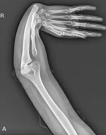
- img1.webp: image
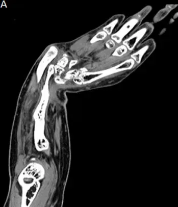
- txt: text

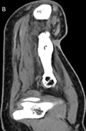
tree (truncated):
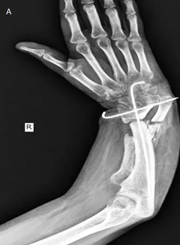
```
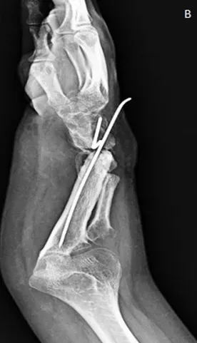
.stats.json
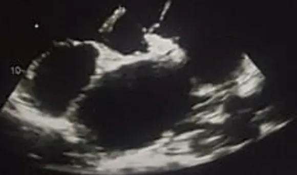
part-000000.tar
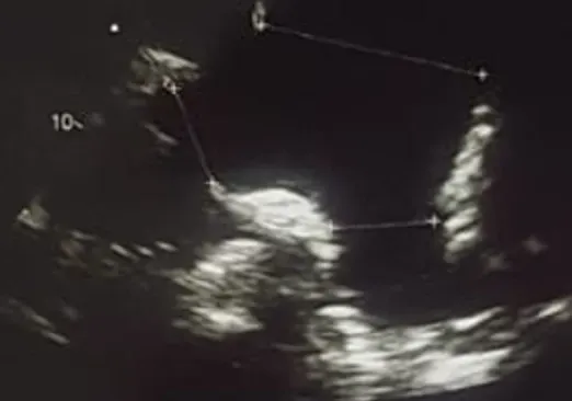
part-000001.tar
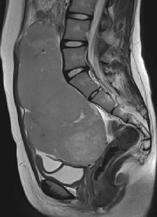
part-000002.tar
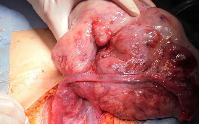
part-000003.tar
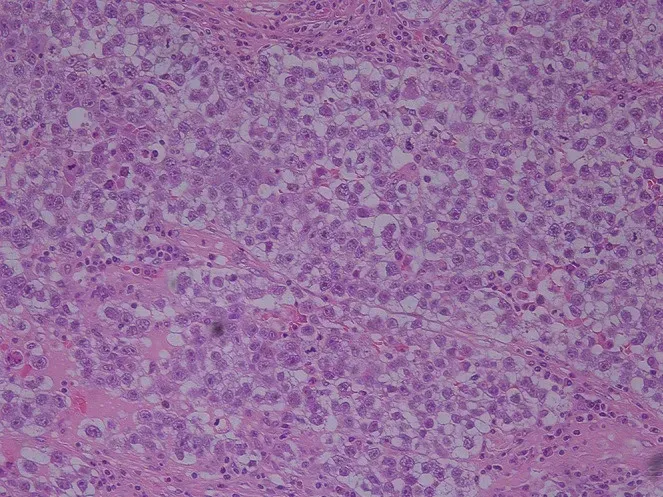
part-000004.tar
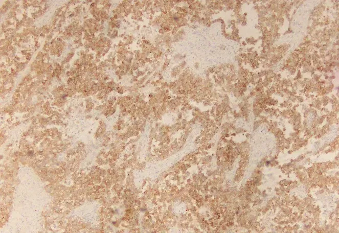
part-000005.tar
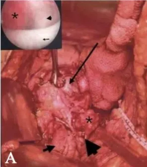
part-000006.tar
part-000007.tar
part-000008.tar
part-000009.tar
part-000010.tar
part-000011.tar
part-000012.tar
part-000013.tar
part-000014.tar
part-000015.tar
part-000016.tar
sample/multicare_images__00000000.img1.webp
sample/multicare_images__00000000.txt
sample/multicare_images__00000001.img1.webp
sample/multicare_images__00000001.txt
sample/multicare_images__00000002.img1.webp
sample/multicare_images__00000002.txt
sample/multicare_images__00000003.img1.webp
... (+288 more)
```
samples
| # | key | img1.webp | txt |
|---|---|---|---|
1 |
multicare_images__00000000 |
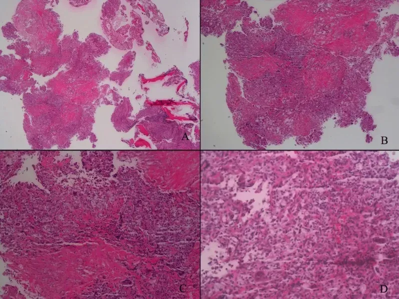
|
<|img1|> Pathological result. |
2 |
multicare_images__00000001 |
|
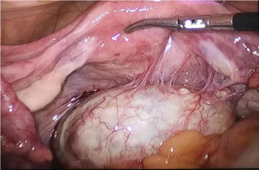
<|img1|> Intraoperative exploration revealed a teratoma in the utero-rectal fossa, adherent to the posterior uterine wall, right adnexa, and mesentery (A). |
3 |
multicare_images__00000002 |
|
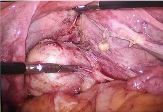
<|img1|> The teratoma was disconnected from the posterior uterine wall, right adnexa, and mesentery (B). |
4 |
multicare_images__00000003 |
|
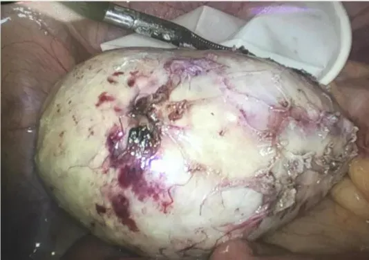
<|img1|> Gross observation of the specimen (C). |
5 |
multicare_images__00000004 |
|
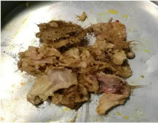
<|img1|> The specimen contains hair, bone, and tooth-like tissues (A). |
6 |
multicare_images__00000005 |
|
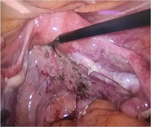
<|img1|> The pelvis and bilateral adnexal areas after complete resection of the teratoma showed no obvious abnormality (B). |
7 |
multicare_images__00000006 |
|
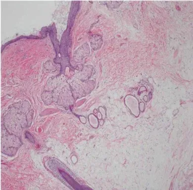
<|img1|> Histological pictures (C). |
8 |
multicare_images__00000007 |
|
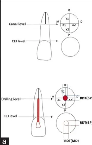
<|img1|> Medical image. |
9 |
multicare_images__00000008 |
|
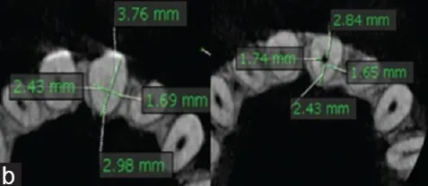
<|img1|> Schematic view of calculation of linear deviation. |
10 |
multicare_images__00000009 |
|
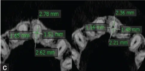
<|img1|> Dentin thickness at CEJ level. Dentin thickness at canal level. CEJ: Cementoenamel junction. |
11 |
multicare_images__00000010 |
|
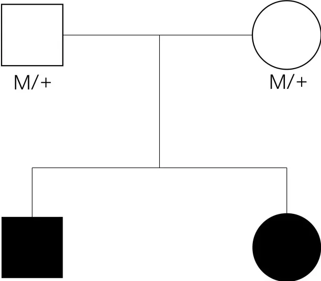
<|img1|> The pedigree of the family. Filled symbol: patient, unfilled symbol: unaffected parents, black arrow: the proband, plus: normal allele, M: mutant allele. |
12 |
multicare_images__00000011 |
|
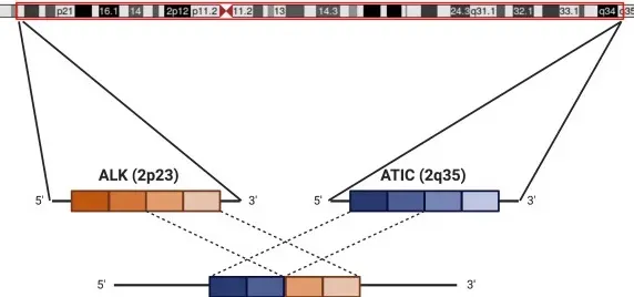
<|img1|> Schematic of ATIC-ALK fusion. Diagram showing chromosome 2 (courtesy of UCSC genome browser; ; with approximate locations of ALK (2p23) and ATIC (2q35). The inversion and subsequent fusion between the 5' end of ATIC to the 3' end of ALK leads to activation of the ALK kinase. For our patient, the fusion is noted specifically between exon 7 of ATIC and exon 20 of ALK. |
13 |
multicare_images__00000012 |
|
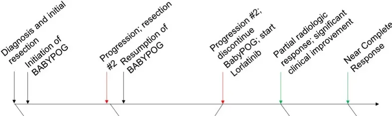
<|img1|> Timeline of major clinical events since diagnosis to present day. |
14 |
multicare_images__00000013 |
|
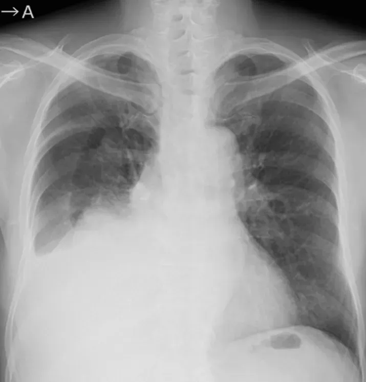
<|img1|> Chest radiograph on admission showing pleural effusion in the right lung. |
15 |
multicare_images__00000014 |
|
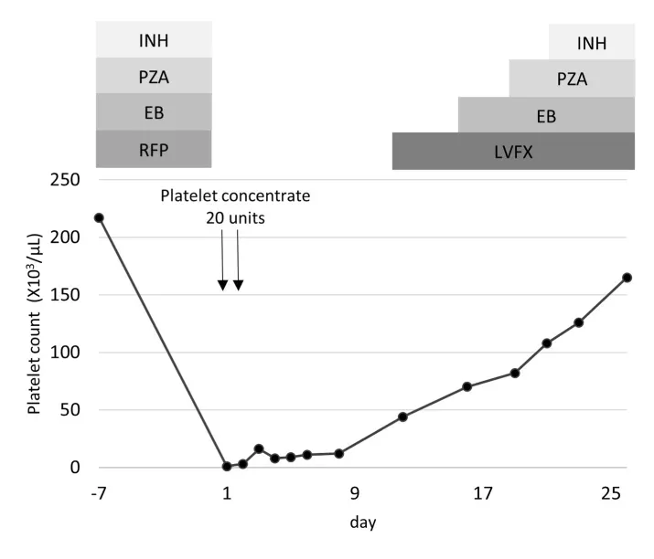
<|img1|> The course of platelet count and treatment. EB: ethambutol, INH: isoniazid, LVFX: levofloxacin, PZA: pyrazinamide, RFP: rifampicin. |
16 |
multicare_images__00000015 |
|
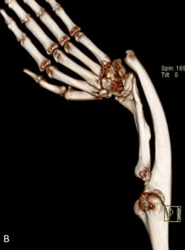
<|img1|> X-ray antero-posterior view. |
17 |
multicare_images__00000016 |
|
<|img1|> Computed tomography 3D reconstruction. Of right forearm showing grossly shortened radius, deformed radius and ulna with radio ulnar dislocation. |
18 |
multicare_images__00000017 |
|
<|img1|> Computed tomography coronal. |
19 |
multicare_images__00000018 |
|
<|img1|> Sagittal. View of right forearm showing non-visualization of the proximal 1/3rd of the radius with the shaft of ulna appearing curved and poorly supporting the wrist joint; muscles of the forearm show disuse atrophy. |
20 |
multicare_images__00000019 |
|
<|img1|> Post-operative (osteotomy of radius and ulna) X-ray antero-posterior. |
21 |
multicare_images__00000020 |
|
<|img1|> Lateral. View of right forearm showing reduction of the deformity using a fibular bone graft and k-wire stabilization. |
22 |
multicare_images__00000021 |
|
<|img1|> transthoracic echocardiography showing ventricular wall defect with dilatation of the four cardiac cavities. |
23 |
multicare_images__00000022 |
|
<|img1|> massive dilatation of the main pulmonary artery measuring 55mm. |
24 |
multicare_images__00000023 |
|
<|img1|> Preoperative pelvic magnetic resonance imaging revealed an 18-cm bifurcated mass with high signal intensity on T2-weighted imaging. |
25 |
multicare_images__00000024 |
|
<|img1|> A small uterus and 18-cm solid tumor in the left ovary were detected intraoperatively. The right fallopian tube was normal, but the right ovary was not detected. |
26 |
multicare_images__00000025 |
|
<|img1|> Histopathological diagnosis was dysgerminoma in the left ovary. Large round cells proliferated like a solid nest with lymphocytic infiltration in the stroma. The cell borders were clear and round with enlarged nuclei, clear cytoplasm, and coarse chromatin staining. |
27 |
multicare_images__00000026 |
|
<|img1|> The ovarian tumor was stained strongly with c-kit. |
28 |
multicare_images__00000027 |
|
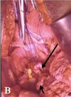
<|img1|> Image showing. Right ovarian cyst (long arrow) densely adhered near the left vaginal vault apex located near the VVF site (arrow head) with right fimbrial herniation (asterisk) into the bladder (short arrow) through the VVF tract. |
29 |
multicare_images__00000028 |
|
<|img1|> Bladder (short arrow), and ,vaginal end (long arrow), post right salpingo-oophorectomy [Cystoscopic view showing papillary frond like mass (asterisk) protruding intravesically (arrow) from VVF site (arrow head). |
30 |
multicare_images__00000029 |
|
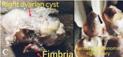
<|img1|> Gross specimen of serous cystadenoma of the right ovary with fimbria. |
31 |
multicare_images__00000030 |
|
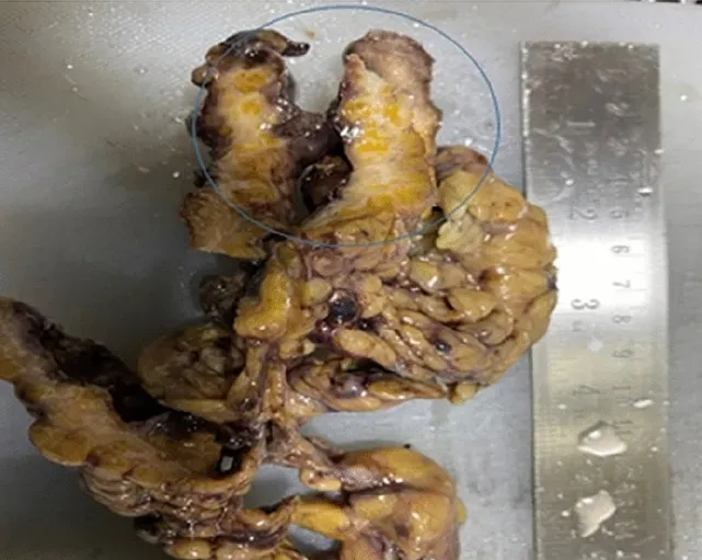
<|img1|> Omentum displaying gray white tumor. |
32 |
multicare_images__00000031 |
|
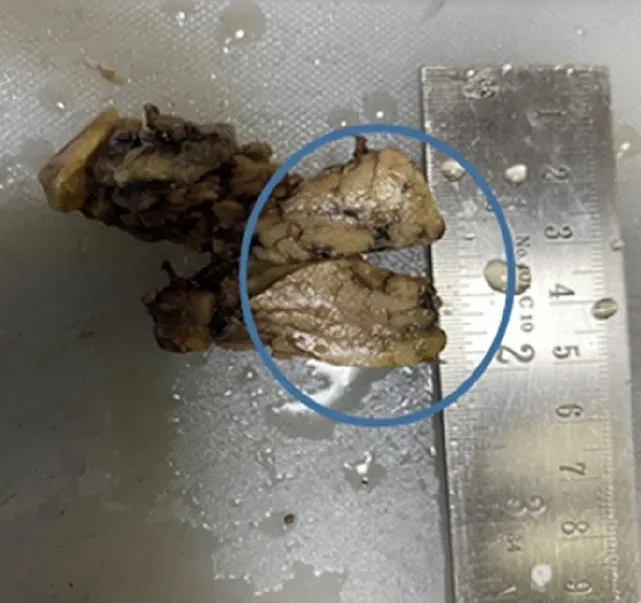
<|img1|> Omentum displaying a gray-tan nodule resembling pancreatic parenchyma. |