| # | key | img1.png | txt |
|---|
1
|
medtrinity_full__00000000
|

|
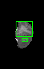
The histopathology image displays a section of breast tissue affected by invasive ductal carcinoma, characterized by a complex arrangement of ductal structures and stromal changes. The image is a microscopic view of the tissue, with the central to upper-middle portion showing a higher density of cells indicative of a higher grade of the carcinoma, while the lower-middle to bottom part of the image reveals a lower grade, suggesting a lower grade tumor. The region of interest, located horizontally in the central to upper-middle and vertically in the middle, exhibits a higher density of cells with a disorganized structure, which is atypical for a grade 2B breast cancer, and contains a mixture of tubule formations and stromal changes. This area's abnormal cellularity and structural disorganization compared to the surrounding tissue could imply a more aggressive disease progression or a higher grade of cancer. The relationship between the region of interest and the surrounding tissue suggests a possible transition from a lower to a higher grade of malignancy, indicating a potential progression of the disease within the tissue.
|
2
|
medtrinity_full__00000001
|
|
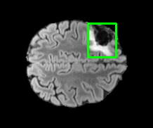
The image is a T2-FLAIR MRI scan of the brain, showing the cerebral hemispheres with the cerebellum not visible in this slice. The region of interest, located in the right-center and upper-middle of the image, occupying approximately 4.8% of the area, exhibits a hyperintense signal, which is indicative of a diffuse glioma. This abnormal signal is surrounded by normal brain tissue, which appears darker on the MRI scan. The region of interest likely represents a pathological change in the brain tissue, which could be exerting pressure on adjacent structures, potentially causing a mass effect or leading to secondary symptoms due to its location and the disruption of normal brain function.
|
3
|
medtrinity_full__00000002
|

|
The image is a histological slide of breast tissue, likely stained with hematoxylin and eosin, showcasing the intricate cellular architecture of the breast tissue, which includes the presence of ductal structures and adipose tissue. The region of interest, located centrally and towards the lower left quadrant of the image, displays a cluster of cells with a high nuclear to cytoplasmic ratio and a high nucleus-to-cytoplasmic ratio, suggesting a high degree of cellular activity and potential malignancy. This area, which is distinct from the surrounding tissue, exhibits a different cellular composition, possibly indicating a more aggressive form of breast cancer. The abnormal cells within this region, characterized by a higher density of irregularly shaped and sized cells with prominent nucleoli and a disorganized structure, could be indicative of a more advanced stage of disease progression. The relationship between this region and the surrounding tissue could suggest that the disease is localized and may be affecting or arising from the ductal structures, potentially impacting adjacent tissues.
|
4
|
medtrinity_full__00000003
|

|
The histopathology image displays a section of breast tissue affected by a carcinoma, with the region of interest located in the central to upper middle part of the image, which is characterized by a dense, irregularly shaped cluster of cells with varying sizes of nuclei and a high nuclear to cytoplasmic ratio, suggesting a high degree of cellular activity indicative of a high-grade malignancy. The region of interest, situated in the central to upper middle area of the image, shows a notable difference in cellular morphology compared to the surrounding tissue, with the affected area displaying a higher density of irregularly shaped and sized nuclei and a disorganized structure, which is not typical of the normal lobular architecture usually seen in benign lesions. This area's abnormal appearance, which includes a mix of cell types such as myoepithelial cells and lymphocytes, may indicate a more aggressive form of cancer, potentially an invasive ductal carcinoma. The abnormal cells within this region suggest a malignant process, which could be influencing or resulting from the surrounding tissue's condition, indicating a possible invasive behavior of the disease that may be affecting or arising from the ductal structures in the breast tissue.
|
5
|
medtrinity_full__00000004
|

|
The image is a histopathology (HP) scan of the brain, showing a cross-sectional view with various shades of gray indicating different tissue densities and compositions. A region of interest is located at left-center part of the image horizontally and middle part of image vertically, occupying approximately 40.5% of the total area. The region of interest, which is highlighted, exhibits an abnormality that differs in appearance from the surrounding brain tissue, potentially indicating the presence of an invasive ductal carcinoma. This abnormal area is characterized by a distinct texture and possibly a difference in size or color intensity, suggesting a pathological change. The affected area's relationship to the surrounding brain tissue could imply a localized pathological process that may be impacting or being impacted by adjacent structures, potentially altering their function or being a result of their dysfunction. The abnormality's proximity to critical brain regions could suggest a potential influence on or from those areas, although the exact nature of this relationship would require further clinical correlation.
|
6
|
medtrinity_full__00000005
|
|
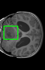
The image is a T1-Post-contrast MRI scan of the brain, showing a detailed view of the brain's structures. The region of interest, located left-center and in the middle of the image, occupies approximately 5.8% of the area and appears to contain an enhancing tumor with a cystic component. This region is characterized by an abnormal signal intensity compared to the surrounding brain tissue, which may indicate the presence of a tumor. The region of interest's proximity to other brain structures suggests a potential relationship, where the tumor could be affecting or displacing adjacent tissues. The abnormal area within the region of interest, when compared to the rest of the brain tissue, could be indicative of a pathological process such as a glioblastoma, which is known to have both enhancing and non-enhancing components, as well as a cystic nature on MRI scans.
|
7
|
medtrinity_full__00000006
|
|
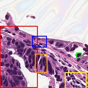
The histological image displays a section of tissue that is likely from the stomach, as indicated by the presence of glandular structures and the absence of signet ring cell hyperplasia, with the region of interest located horizontally in the center and vertically in the lower-middle, occupying approximately 2.8% of the area. Within this region, there are noticeable differences in cellular morphology and staining patterns, such as altered color and texture, which may suggest pathological changes. The affected area is characterized by a cluster of cells with a lower nucleus-to-cytoplasm ratio and a higher nucleus-to-cytoplasm ratio, which could indicate a reduction in cellular activity or a different phase of cellular activity. This region's proximity to other cellular structures suggests a potential relationship, possibly indicating that the changes within the region of interest could be a localized manifestation of the disease process affecting the stomach, with the possibility of the surrounding tissue being either a source of the disease or being secondarily affected by the pathological process occurring within the central area.
|
8
|
medtrinity_full__00000007
|
|
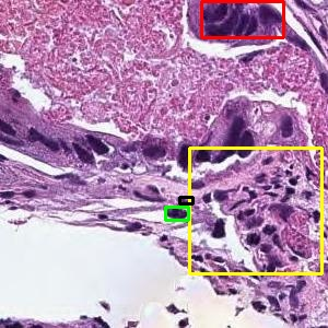
The image is a histological slide of lung tissue, likely from a bronchus, showing a detailed view of the bronchus epithelium with a focus on the bronchial wall, which is located in the right-center horizontally and lower-middle vertically, occupying approximately 0.3% of the area. The region of interest, situated in the central to lower-middle part of the image, displays abnormal tissue characteristics, including changes in color and texture that suggest a moderately differentiated adenocarcinoma, with the affected area showing a mix of papillary structures and a dense inflammatory response. The unusual features within this region, which is not typical of normal lung tissue, such as altered glandular structures and the presence of necrotic cells, could indicate a pathological condition. The abnormal tissue in the region of interest, characterized by a cluster of nuclei that are indicative of a disease process, potentially a moderately differentiated adenocarcinoma. The affected area's proximity to the bronchus suggests a possible relationship with the surrounding lung tissue, which may be either a result of the disease spreading from or to the bronchus or a localized response to an adjacent area, although the exact nature of this relationship cannot be determined without further clinical correlation.
|
9
|
medtrinity_full__00000008
|
|
The image is a T2-FLAIR MRI scan showing a cross-sectional view of the brain, with the region of interest located centrally and occupying approximately 0.6% of the area. This region exhibits abnormal signal intensity, which is indicative of a pathological condition, likely representing necrosis, edema, or an area of the brain that is jointly affected by the disease. The region of interest's abnormal appearance suggests tissue damage, which could be related to the surrounding brain structures either as a consequence of the same pathological process or as a potential cause of secondary effects due to its central location.
|
10
|
medtrinity_full__00000009
|

|
The histopathology image displays a section of breast tissue, where the central upper-middle region, occupying approximately 10.1% of the area, exhibits a cluster of necrosis nuclei. These nuclei are characterized by their distinct appearance, which differs from the surrounding tissue in terms of color and texture, suggesting a pathological change. The necrotic area is surrounded by other cellular structures, which may be either contributing to the disease process or being affected by it, indicating a potential invasive ductal carcinoma within the breast tissue.
|
11
|
medtrinity_full__00000010
|
|
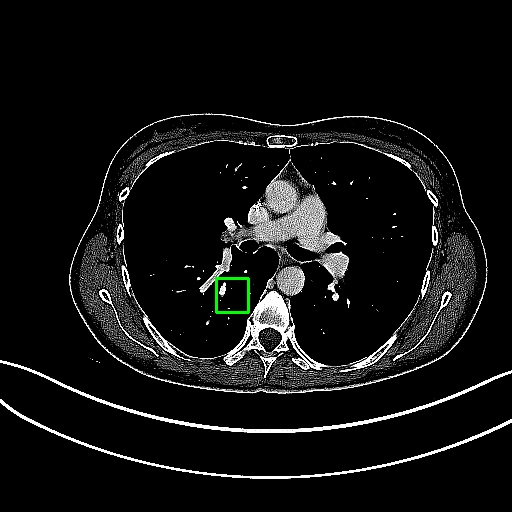
The image is a transverse section of a computed tomography (CT) scan of the chest, showing the lungs, heart, and part of the spine. The heart is centrally located with the lungs on either side, and the spine is visible posteriorly. The region of interest, located centrally and horizontally in the middle of the image, encompasses a small area of the lung tissue. Within this region, there is an abnormality that differs in texture and possibly density from the surrounding lung parenchyma, which could indicate a lesion. This lesion's proximity to the heart and major blood vessels suggests a potential relationship, as diseases of the lung can sometimes affect nearby structures or be influenced by them due to their close anatomical association. However, without further clinical context, it is not possible to definitively determine the nature of this relationship.
|
12
|
medtrinity_full__00000011
|
|
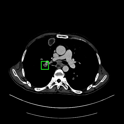
The image is a transverse section of a computed tomography (CT) scan of the chest, showing the lungs, heart, and part of the spine. The heart is centrally located, with the lungs on either side and the spine visible posteriorly. The region of interest, located in the left-center of the image and occupying approximately 0.4% of the area, shows an abnormality within the lung tissue. This abnormality is characterized by a difference in texture and possibly density compared to the surrounding lung parenchyma, which may indicate the presence of a lesion. The lesion's proximity to the heart and major blood vessels could suggest a relationship where the lesion might affect or be affected by the adjacent structures, such as the spread of inflammation or malignancy from nearby tissues. However, without additional clinical information, the exact nature of this relationship cannot be determined.
|
13
|
medtrinity_full__00000012
|
|
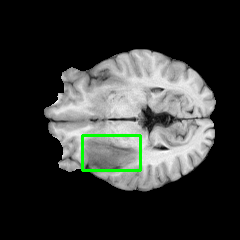
The image is a T1-Post-contrast MRI scan of the brain, showing a detailed view of the brain's structures. The region of interest, located centrally and in the lower-middle part of the image, occupies approximately 3.5% of the area and exhibits abnormal signal intensity, which is indicative of a brain tumor with associated edema. This region's unusual appearance, characterized by differences in color and texture compared to the surrounding brain tissue, suggests the presence of a pathological process. The abnormal area is likely to be affecting or being affected by adjacent brain structures due to its central location, which could imply an influence on nearby functional areas of the brain or the presence of edema that often accompanies such tumors.
|
14
|
medtrinity_full__00000013
|
|
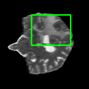
The image is a T2-weighted MRI scan of the brain, showing a cross-sectional view with various brain structures visible. The region of interest, located centrally and in the upper-middle area of the image, occupies approximately 15.0% of the area and displays an abnormality indicative of a diffuse glioma. This abnormality is characterized by a difference in signal intensity compared to the surrounding brain tissue, which may suggest the presence of a tumor. The region of interest is situated near critical brain structures, and its abnormal appearance could potentially affect or be affected by these adjacent areas. The relationship between the content in the region of interest and other regions could be that the tumor is causing a mass effect or displacement of nearby brain structures, which is a common occurrence with intracranial tumors.
|
15
|
medtrinity_full__00000014
|
|
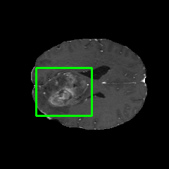
The image is a magnetic resonance imaging (MRI) scan showing a cross-sectional view of the brain, with the focus on the left-center, middle part of the brain. The region of interest, occupying approximately 9.3% of the area, exhibits abnormal tissue characteristics indicative of a diffuse glioma, such as altered signal intensity, which may suggest the presence of necrosis, edema, or an enhancing tumor. This abnormal region is likely to be affecting or being affected by the surrounding brain structures due to its location and the nature of brain gliomas to influence adjacent tissues.
|
16
|
medtrinity_full__00000015
|

|
The image is a magnetic resonance imaging (MRI) scan of the brain, showing a detailed view of the brain's structures. The region of interest, located centrally within the brain tissue, exhibits an abnormality indicative of invasive ductal carcinoma, characterized by a distinct texture and possibly a difference in intensity or color compared to the surrounding brain tissue. This abnormal area could be affecting or be affected by adjacent brain structures due to its central location, potentially influencing nearby tissue or being influenced by it, as the central nervous system is often interconnected with various brain regions. The abnormality's proximity to critical brain structures suggests a possible impact on or from these areas, although the exact relationship depends on the specific brain region involved and the nature of the pathology.
|
17
|
medtrinity_full__00000016
|
|
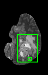
The image is a sagittal section of a magnetic resonance imaging (MRI) scan showing the brain, with the region of interest located in the right-center, lower-middle portion, occupying approximately 0.3% of the area. This region exhibits abnormal signal intensity, which is indicative of a pathological condition, likely representing necrosis, edema, or an enhancing tumor. The region of interest's relative position to other brain structures suggests it could be affecting or be affected by adjacent tissues, potentially disrupting normal brain function due to its pathological nature.
|
18
|
medtrinity_full__00000017
|

|
The histopathology image displays a section of breast tissue, where the central and upper right quadrant shows a cluster of cells with a high nuclear grade, suggesting a more aggressive form of cancer, likely an invasive ductal carcinoma, which is characterized by a higher likelihood of metastasis. The region of interest, located centrally and towards the upper right, exhibits abnormal cell morphology with variations in color and texture that are indicative of malignancy. This area's abnormal cells are in stark contrast to the surrounding tissue, which may be normal breast tissue, implying that the disease could be spreading from this central area to other parts of the body, potentially affecting adjacent tissues or being a result of the progression from a nearby affected area.
|
19
|
medtrinity_full__00000018
|

|
The histopathology image displays a section of breast tissue affected by invasive ductal carcinoma, with the most prominent organ being the breast. The image is a microscopic view of the tissue, where the cells exhibit a high nuclear grade, nuclear pleomorphism with a high mitotic rate and numerous atypical mitotic figures, suggesting a high proliferation rate and aggressive tumor behavior. The region of interest, located centrally and extending towards the lower right quadrant, shows a dense cellular arrangement with a high nuclear grade and mitotic activity, indicative of a more aggressive cancer subtype. This area, characterized by a higher density of irregularly shaped and sized nuclei, is likely to be the primary site of the disease, which may influence the surrounding tissue due to its aggressive nature, potentially leading to further spread of the carcinoma.
|
20
|
medtrinity_full__00000019
|
|
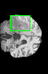
The image is a magnetic resonance imaging (MRI) scan of the brain, showing the cerebral structures without any medical devices present. The region of interest, located centrally and in the upper-middle area of the brain, occupies approximately 8.3% of the image area and displays an abnormality indicative of a diffuse glioma. This abnormality is characterized by a difference in signal intensity compared to the surrounding brain tissue, which may suggest the presence of edema or an invasive tumor. The region of interest's proximity to other cerebral structures could imply a potential impact on or from adjacent areas of the brain, possibly affecting their function or being affected by the pathology present.
|
21
|
medtrinity_full__00000020
|

|
The histopathology image displays a section of breast tissue, where the central to lower middle region exhibits a cluster of cells with a high nuclear to cytoplasmic ratio, indicative of invasive ductal carcinoma. The specific area of interest, characterized by a dense aggregation of small, round, and uniform nuclei, is situated in the central to lower middle part of the image, which is significant as it suggests a higher likelihood of malignancy. This area of interest, with its distinct cellular features, is likely to be the primary site of the carcinoma, influencing the surrounding tissue and potentially representing the origin of the disease.
|
22
|
medtrinity_full__00000021
|

|
The image is a sagittal section of a T2-FLAIR MRI scan, showing the central part of the brain with a focus on the region that includes the corpus callosum and surrounding white matter. The region of interest, located centrally and in the middle of the image, occupies approximately 5.4% of the area and displays a hyperintense signal, which is indicative of a diffuse glioma. This abnormal signal suggests the presence of a tumor that is not enhancing with contrast, which could be related to the surrounding brain tissue either by causing a mass effect or by being affected by the same pathological process that caused the tumor. The relationship between the tumor and adjacent brain structures could imply a potential impact on nearby functional areas, depending on the extent of the tumor's growth and infiltration.
|
23
|
medtrinity_full__00000022
|
|
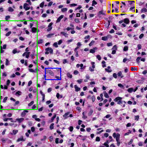
The image is a histopathological slide of brain tissue, likely stained with hematoxylin and eosin, showing the intricate cellular architecture of a glioblastoma, a type of malignant brain tumor. The slide captures a detailed view of the brain's cellular composition, including the presence of tumor cells, normal brain tissue, and possibly reactive astrocytes, with the tumor cells displaying a high degree of pleomorphism and a high mitotic rate. The region of interest, located at the right-center horizontally and at the top vertically, occupying approximately 0.8% of the area, exhibits abnormal features such as irregular cell shapes and sizes, and a disorganized structure, which are indicative of glioblastoma. This area is distinct from the surrounding tissue, which may be either unaffected or less affected by the disease, suggesting that the region of interest could be a focal point of the disease's manifestation, potentially influencing or being influenced by the adjacent tissue due to its aggressive nature.
|
24
|
medtrinity_full__00000023
|
|
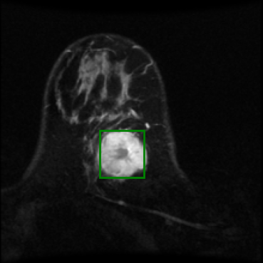
The image is a magnetic resonance imaging (MRI) scan of the brain, showing a detailed view of the brain's structures. The region of interest, located centrally and in the middle of the image, occupies approximately 3.0% of the area and exhibits an abnormality indicative of brain cancer. This abnormality is characterized by a difference in texture and contrast compared to the surrounding brain tissue, suggesting a possible mass effect or pathological change. The region of interest's proximity to other brain structures may imply a potential impact on or from these adjacent areas, possibly affecting their function or being influenced by surrounding tissue characteristics.
|
25
|
medtrinity_full__00000024
|
|
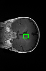
The image is a T1-weighted MRI scan of the brain, showing the cerebral hemispheres, ventricles, and other intracranial structures. The region of interest, located centrally and in the middle of the image, appears to have a different signal intensity compared to the surrounding brain tissue, which may indicate a non-enhancing tumor. This abnormal area is likely related to the surrounding brain structures, potentially affecting or being affected by them due to its central location, which is critical for various brain functions.
|
26
|
medtrinity_full__00000025
|
|
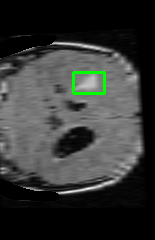
The image is a T2-FLAIR MRI scan of the brain, showing a non-enhancing tumor located centrally and in the upper-middle area, occupying approximately 1.8% of the image area. The region of interest, indicative of the disease, is characterized by an abnormal signal intensity compared to the surrounding brain tissue. This region's relative position to other brain structures suggests it could be affecting or be affected by adjacent areas due to its central location, which is critical for various brain functions.
|
27
|
medtrinity_full__00000026
|
|
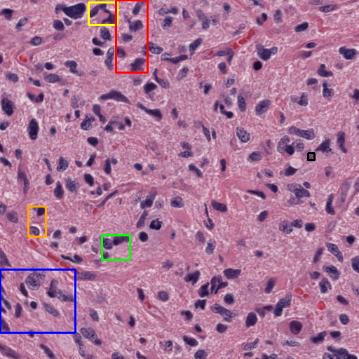
The histopathology image displays a section of brain tissue, where the region of interest, located horizontally on the left and vertically in the lower-middle, occupying an area ratio of 0.4%, exhibits abnormal features. Within this area, there are cells with large, irregular nuclei and prominent nucleoli, which are indicative of a glioblastoma, a malignant brain tumor. These abnormal cells are surrounded by more typical brain tissue, suggesting that the tumor may be infiltrating from the region of interest into adjacent areas, potentially disrupting normal brain function and structure.
|
28
|
medtrinity_full__00000027
|
|
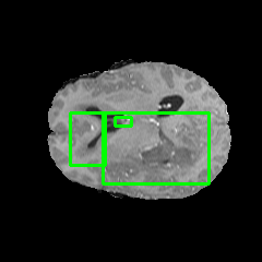
The image is a T1-weighted MRI scan of the brain, showing a detailed view of the brain's structures. The region of interest, located centrally and in the middle of the image, occupies approximately 0.2% of the area and exhibits an abnormality indicative of a diffuse glioma. This abnormality is characterized by a difference in texture and possibly color compared to the surrounding brain tissue. The affected area's proximity to critical brain structures suggests a potential impact on or from these regions, possibly indicating an interrelationship where the glioma could be exerting pressure on or infiltrating nearby tissues.
|
29
|
medtrinity_full__00000028
|
|
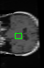
The image is a post-treatment MRI scan of the brain, showing a non-enhancing tumor located centrally and in the middle of the brain, occupying approximately 1.2% of the area. The region of interest, indicative of the disease, is characterized by an abnormality in tissue appearance, which may present as a difference in color or texture compared to the surrounding brain tissue. This abnormal region could potentially be affecting or be affected by adjacent brain structures due to its central location, which is critical for various brain functions. The relationship between this region and its surroundings could suggest an impact on or from nearby neural pathways or brain regions.
|
30
|
medtrinity_full__00000029
|
|
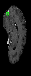
The image is a sagittal section of a T1-weighted MRI scan, showing the cerebral hemispheres, ventricles, and other intracranial structures. The region of interest, located at the top left-center of the image, occupies approximately 0.2% of the area and is characterized by an abnormal signal intensity, which is indicative of a diffuse glioma. This region is situated adjacent to the lateral ventricles and appears to have a different texture and signal intensity compared to the surrounding brain tissue, suggesting a pathological process. The abnormal area could be exerting pressure on adjacent structures or could be a result of infiltration by the disease process, potentially affecting nearby brain functions due to its proximity to critical areas.
|
31
|
medtrinity_full__00000030
|

|
The image is a magnetic resonance imaging (MRI) scan of the brain, showing the intricate structures within the cranial cavity. The region of interest, located centrally within the brain tissue, exhibits abnormal signal characteristics that are indicative of an invasive ductal carcinoma. This area is surrounded by normal brain parenchyma, which provides a stark contrast to the irregularity of the affected tissue. The abnormal region likely represents a pathological change, possibly due to the presence of a tumor that has altered the normal brain architecture and could be exerting pressure on or displacing adjacent brain structures. The relationship between the region of interest and the surrounding brain tissue suggests that the tumor may be affecting nearby areas either by direct invasion or through secondary effects such as edema or mass effect.
|
32
|
medtrinity_full__00000031
|
|
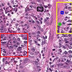
The histological image displays a section of tissue that is likely from the stomach, showing a moderately differentiated tumor with a mix of glandular and non-glandular areas, indicative of a moderately differentiated tubular adenocarcinoma. The region of interest, located at the top right of the image, occupying approximately 0.1% of the area, exhibits a cluster of cells with a high nuclear-cytoplasmic ratio and a disorganized structure, contrasting with the surrounding tissue. This area, which is unusual due to its densely packed cells with prominent nuclei and lack of glandular formation, suggests a more aggressive tumor type. The affected area's proximity to other tissue structures could imply that the disease may be spreading from this highly cellular region to adjacent areas, potentially indicating an invasive growth pattern of the adenocarcinoma.
|